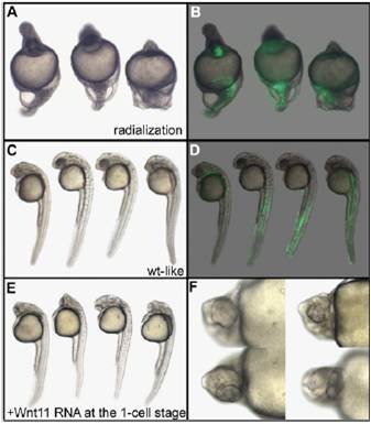
Fig. S6 Phenotype and localization of the injected clone of cells in nocodazole-treated embryos injected with Wnt11 and GFP mRNAs. Embryos displaying a radialized phenotype after treatment with nocodazole and injection of 400 pg of Wnt11 and 100 pg of GFP mRNAs (A and B) demonstrate that Wnt11 is unable to rescue the lack of stimulation of blastomeres by dorsal determinants. Embryos displaying a wild-type–like phenotype (C and D) express the GFP in lateral and ventral (muscle and blood) but not in axial tissues (notochord and hatching gland). (E and F) Embryos injected with 400 pg of Wnt11 mRNA at the one-cell stage display cyclopia, demonstrating that the Wnt11 mRNA used is fully active. Embryos are 30 h old, anterior to the Top in lateral view (A–E), and seen in high magnification in ventral view (F).
Image
Figure Caption
Acknowledgments
This image is the copyrighted work of the attributed author or publisher, and
ZFIN has permission only to display this image to its users.
Additional permissions should be obtained from the applicable author or publisher of the image.
Full text @ Proc. Natl. Acad. Sci. USA

