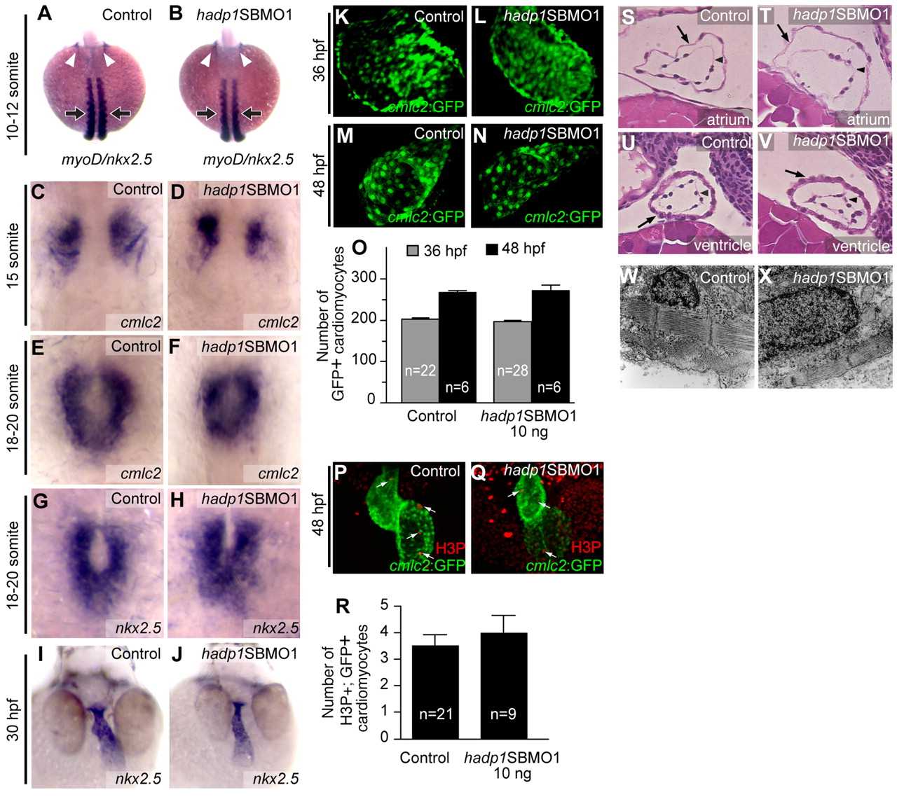
Fig. 4
Cardiogenesis is not altered in hadp1 morphants. (A,B) Early specification of the heart field was analyzed by in situ hybridization for nkx2.5 at the 10- to 12-somite stage. Arrowheads indicate nkx2.5 expression. Embryos were stage-matched according to somite number as determined by myoD staining (indicated by the arrows). (C,D) Specification was also examined by in situ hybridization for cmcl2 at the 15-somite stage. (E–H) Migration of cardiomyocytes to the midline was examined in 18- to 20-somite-stage embryos following loss of hadp1 by in situ hybridization for cmlc2 (E,F) and nkx2.5 (G,H). (I,J) Heart-tube elongation and extension was assessed in 30-hpf control and hadp1 morphant embryos by in situ hybridization for nkx2.5. (K–N) Direct immunofluorescence (K,L) and confocal microscopy (M,N) of cmlc2:GFP hearts from 36 hpf (K,L) and 48 hpf (M,N) control and hadp1 morphant embryos. (O) The number of GFP+ cells was quantified. (P,Q) Confocal analysis of vibratome sectioned hearts following immunostaining for H3P and GFP in 48-hpf control (P) and hadp1 morphant (Q) embryos. (R) The number of double-positive (H3P+, GFP+; i.e. proliferating cardiomyocytes) was quantified. (S–V) Coronal sections through the heart at 48 hpf, stained with hematoxylin and eosin, anterior to the top. Arrow and arrowhead indicate myocardium and endocardium, respectively. (S,T) Comparison of sections through the atrium of control (S) and hadp1 morphant (T) embryos. (U,V) Comparison of sections through the ventricle of control (U) and hadp1 morphant (V) embryos. (W,X) Coronal section at 48 hpf through cardiomyocytes viewed by TEM. Cardiomyocytes in control (W) and hadp1 morphant (X) ventricles contain organized myofibrillar arrays.

