
|
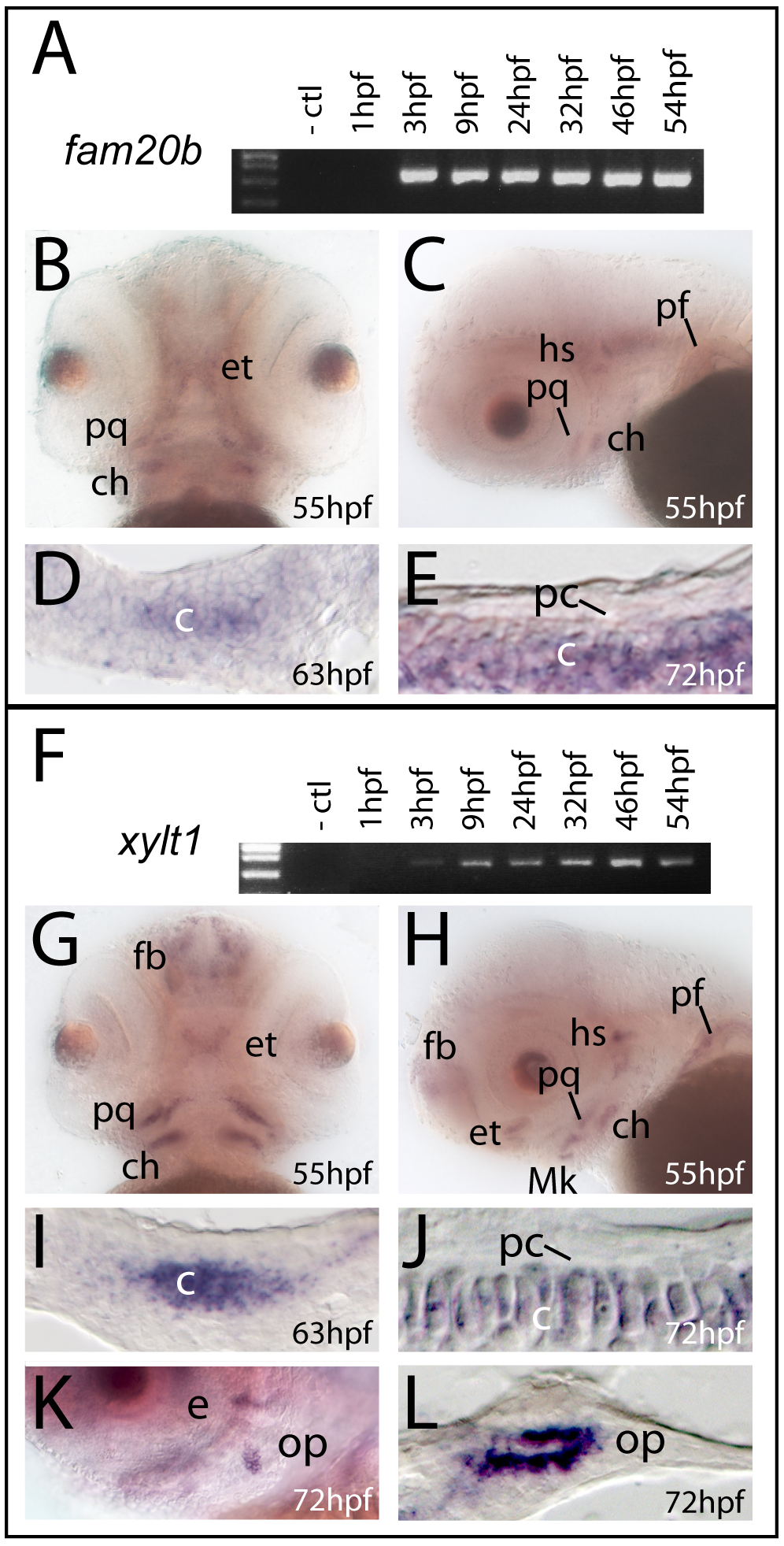
Fig. 4
fam20b and xylt1 are expressed in chondrocytes, but not in perichondrium.
A,F, RT-PCR on wild-type extracts at ages indicated; B,C,G,H,K, whole-mount and D,E,I,J,L section in situ hybridization on wild-type embryos. RT-PCR demonstrated transcripts for fam20b (A) and xylt1 (F) from 3 hpf through 54 hpf. Frontal and lateral views of embryos stained by whole-mount in situ hybridization revealed transcripts for fam20b (B,C) and xylt1 (G,H) in developing cartilage elements of the craniofacial and pectoral fin skeletons at 55 hpf. In addition, there was diffuse expression of fam20b in the brain and specific xylt1 expression in the forebrain (fb). Horizontal section in situ hybridization localized transcripts for fam20b and xylt1 to developing chondrocytes (c) of the ceratohyal at 63 hpf (D,I) and 72 hpf (E,J), but no expression in perichondrium (pc) was detected. Lateral view of whole-mount (K) and horizontal section (L) in situ hybridization revealed expression of xylt1 in osteoblasts of the opercle (op) at 72 hpf. Abbreviations: -ctl = negative control; c = chondrocytes; ch = ceratohyal; e = eye; et = ethmoid; fb = forebrain; hpf = hours post-fertilization; hs = hyosymplectic; Mk = Meckel′s; op = opercle; pc = perichondrium; pf = pectoral fin; pq = palatoquadrate.