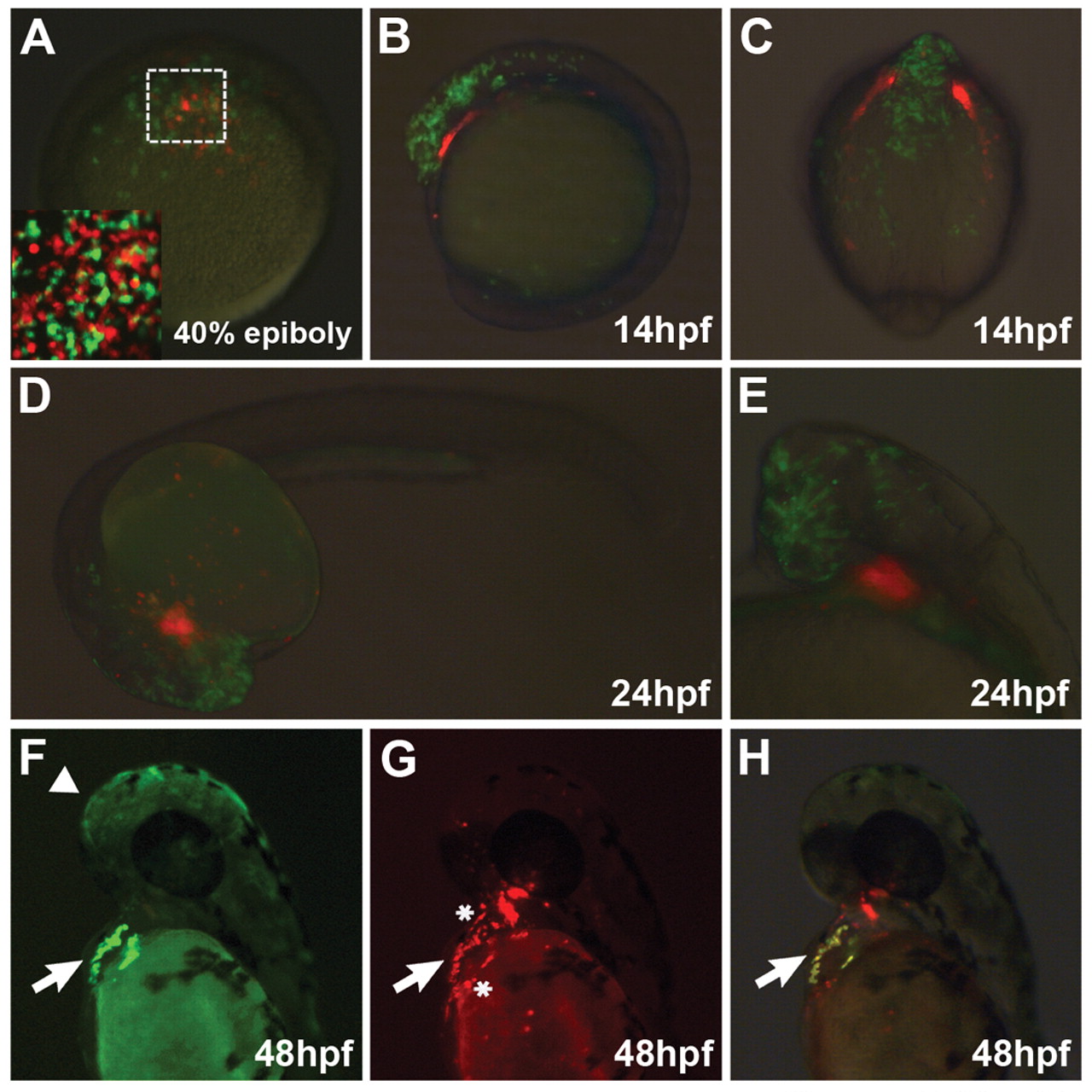
Fig. 5
Cell-autonomous migration of gata5/smarcd3b-expressing cells to the heart. (A-E) Donor cells overexpressing gata5/smarcd3b (red) and wild-type cells marked by rhodamine green were co-transplanted to the animal pole of a wild-type 4 hpf host embryo (A). Boxed area in A is shown at higher magnification in the inset, showing that wild-type and cBAF donor cells are uniformly dispersed in the animal cap following transplantation. (F-H) Co-transplants where gata5/smarcd3b-expressing donor cells carry the myl7:EGFP transgene. Faint rhodamine green signal is evident in the head (arrowhead), whereas stronger EGFP is seen in the heart (arrow). (A) Dorsal view; (B,F-H) lateral views with anterior towards the top; (C) dorsal view onto head; (D,E) lateral views with anterior towards left. Asterisks in G denotes rhodamine-positive cells inside and adjacent to the heart that do not express EGFP.

