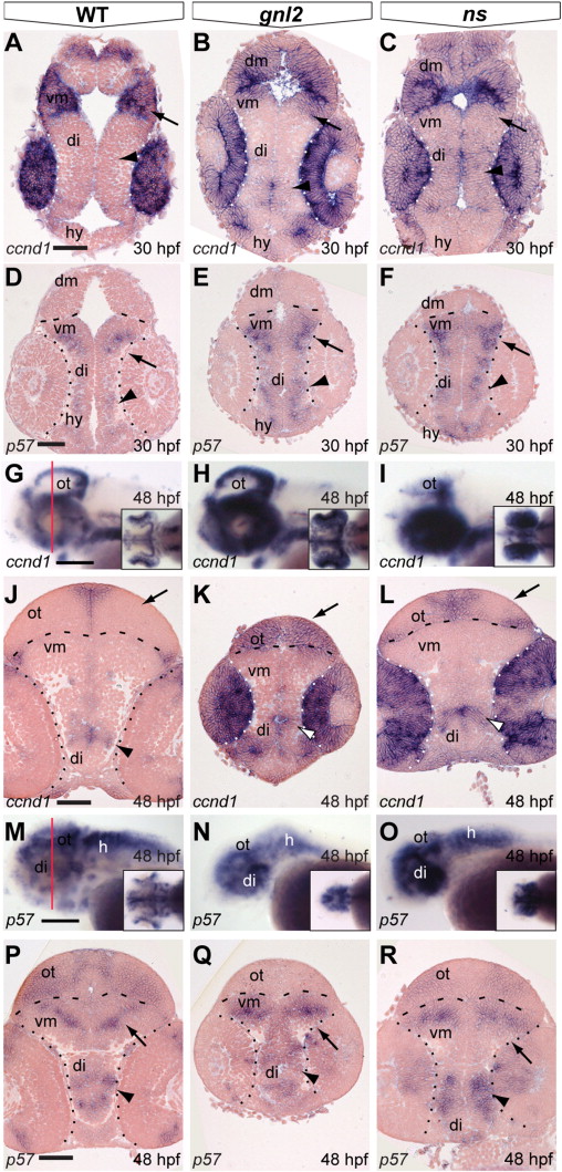
Fig. S4
Differential expression of ccnd1 and p57 in the gnl2 and ns brain and retina. (A–C) Coronal sections showing expression of ccnd1 is expanded in the gnl2 (n = 5; B) and ns (n = 4; C) midbrain (arrow) and diencephalon (arrowhead) as compared to WT embryos at 30 hpf. Dotted lines indicate boundary between retina and brain. (D–F) Expression of the cell cycle inhibitor p57kip2 in the retina and brain at 30 hpf. At 30 hpf, there is no p57kip2 expression in the gnl2 (n = 4; E) and ns mutant retina (n = 4; F), whereas p57kip2 is expanded in the mutant diencephalon (arrowhead) and ventral midbrain (arrow) as compared to WT embryos (D). Dashed lines indicate the boundary between dorsal and ventral midbrain. Dotted lines indicate the boundary between retina and brain. Dorsal up. (G–I) Whole-mount labeling of ccnd1 transcript shows upregulation in the gnl2 (n = 9; H) and ns (n = 9; I) optic tectum, cerebellum and eye at 48 hpf. Lateral view, dorsal up. Insets show dorsal view. Red line in (G) indicates level of sectioning in (J–L). (J–L) Coronal sections show expansion of ccnd1 in the gnl2 (K) and ns (L) mutant optic tectum (arrow) and diencephalon (arrowhead) and retina as compared to WT embryos (J). (M and N) Whole-mount view of p57kip2 expression shows upregulation in the ventral brain of gnl2 (n = 12; N) and ns (n = 13; O) embryos as compared to WTs (M). Lateral view, dorsal is up. Insets show dorsal view. Red line in (M) indicates level of sectioning of (P–R). (P–R) Coronal sections showing expanded p57kip2 expression in the ventral midbrain (arrow) and parts of the diencephalon (arrowheads) of gnl2 (Q) and ns (R) mutants. di, diencephalon; dm, dorsal midbrain; hy, hypothalamus; ot, optic tectum; vm, ventral midbrain. Scalebar 50 μm.
Reprinted from Developmental Biology, 355(2), Paridaen, J.T., Janson, E., Utami, K.H., Pereboom, T.C., Essers, P.B., van Rooijen, C., Zivkovic, D., and Macinnes, A.W., The nucleolar GTP-binding proteins Gnl2 and nucleostemin are required for retinal neurogenesis in developing zebrafish, 286-301, Copyright (2011) with permission from Elsevier. Full text @ Dev. Biol.

