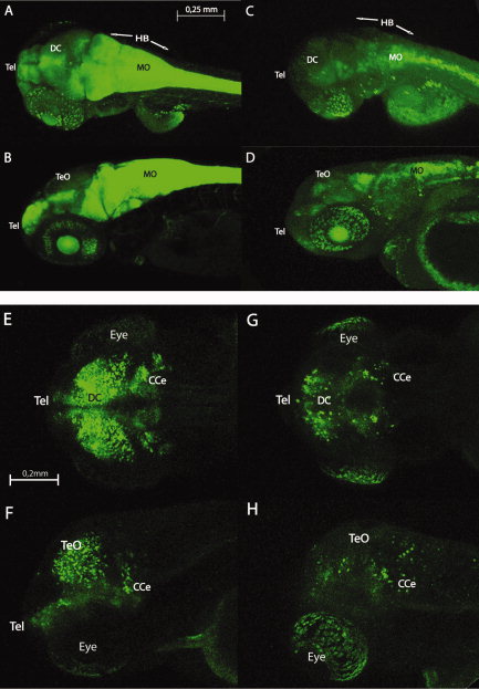
Fig. 10
A–H: Confocal live whole-mount images of Gfap:GFP (upper panel) and Dlx5-6:GFP transgenic zebrafish brain (lower panel) in wild-type (left side) and mibhi904 mutant (right side) at 3 days postfertilization (dpf): dorsal views (A,C,E,G) and lateral views (B,D,F,H). The transgenic line for the glial fibrillary acidic protein (GFAP) drove expression (shown as green fluorescence) initially in the telencephalon and then in the eye and midbrain. Gfap:GFP is also highly prominent in hindbrain in wild-type (WT)siblings with a weak expression pattern in mibhi904 mutants. Dlx5-6:GFP in mibhi904 mutants is reduced with only few cells showing expression in diencephalon, eyes, and in the cerebellar region. Images of live zebrafish were obtained using a Leica SP5 confocal scanning fluorescence microscope. Abbreviations as above. Scale bars = 0.25 mm in A–D, and 0.2 mm in E–H.

