Fig. 4
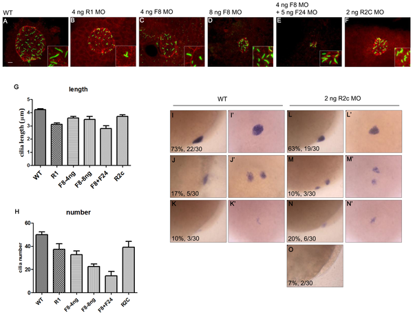
The cilia length was disrupted in fgfr2c morphants.
The KV and cilia were labeled with antibodies against aPKC (red) and acetylated tubulin (green), respectively, at 10 somite-stage embryos (A~F). The cilia length was reduced in fgfr1, fgf8 morphants and fgf8/fgf24 double morphants compared to wild type embryos (A~E, G). The cilia length was also reduced in fgfr2c morphants (F~G). The cilia number was reduced in fgfr1, fgf8 morphants and fgf8/fgf24 double morphants compared to wild type embryos (A~E, H). In fgfr2c morphants, the cilia number was not significantly reduced (F, H, 39.2±4.9, P = 0.0725). Various expression patterns of foxj1a were detected in wild type embryos (lateral view, I~K; dorsal view, I2~K2) and fgfr2c morphants (lateral view, L~O; dorsal view, L2~N2) at 90% epiboly. Error bar, s.e.m. Scale bar: 10 μm.

