
Image
|
Figure Caption
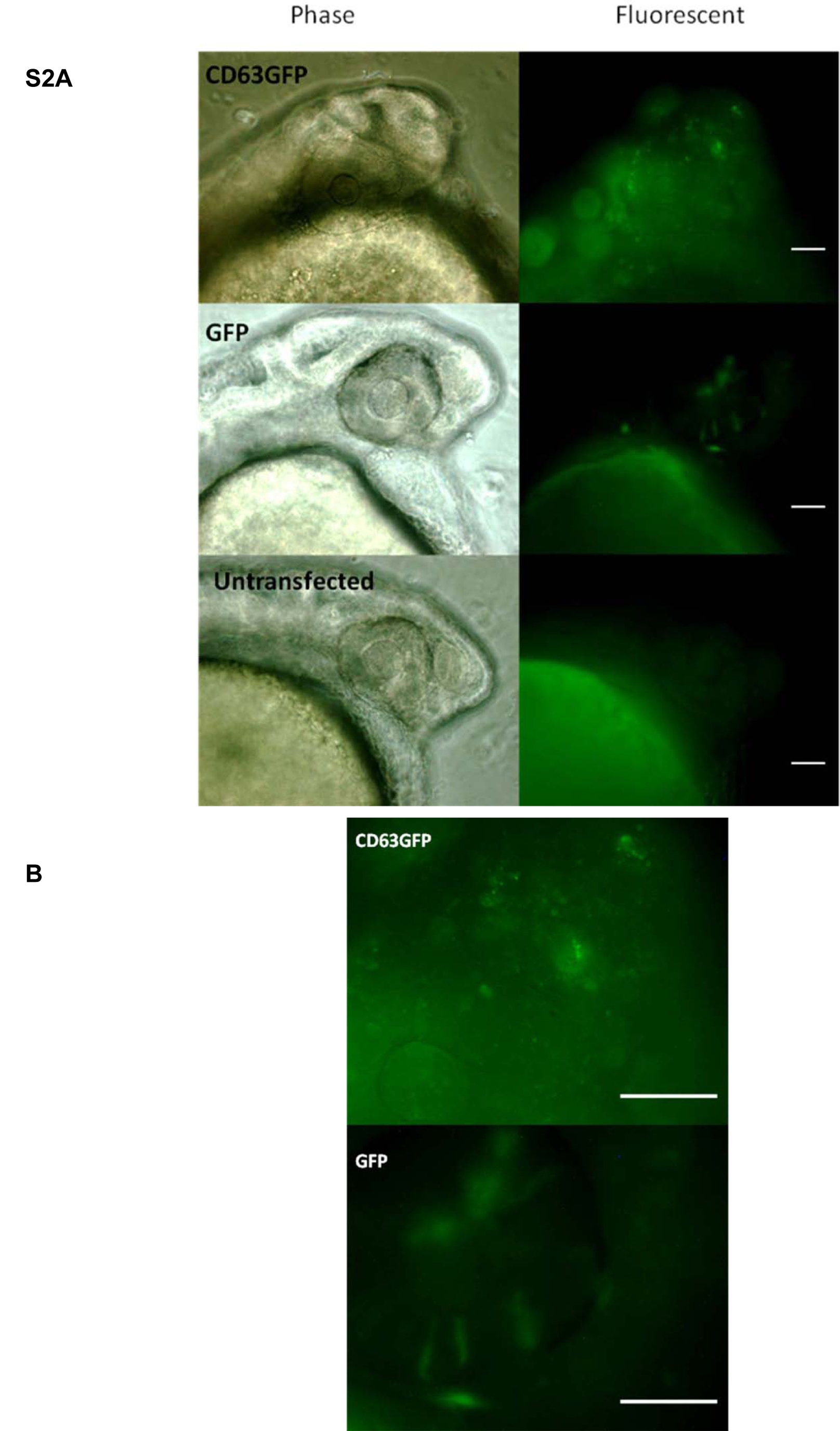
Fig. S2
A. Localisation of Cd63GFP in 24 hr LWT embryos injected with buffer or plasmid DNA encoding Cd63GFP or EGFP, as indicated. Scale bars = 52 μm. B. Zoom of area of fluorescent Cd63GFP and GFP images in S2 A. Scale bars = 52 μm.
Acknowledgments
This image is the copyrighted work of the attributed author or publisher, and
ZFIN has permission only to display this image to its users.
Additional permissions should be obtained from the applicable author or publisher of the image.
Full text @ PLoS One