
|
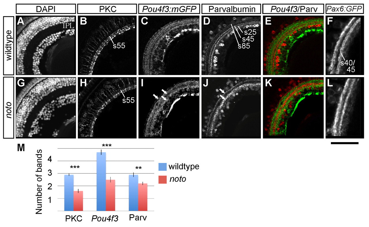
Fig. 1
Fine neurite targeting is disrupted in the 5 dpf noto mutant inner plexiform layer (IPL). (A-L) Representative sections of wild-type (A-F) and mutant (G-L) zebrafish retina. DAPI nuclear stain shows the cellular organization of the retina, including location and width of the IPL (A,G). Protein kinase C (PKC)+ bipolar cells (BCs) project terminals to three IPL sublaminae in wild type (B); in the mutant, outermost sublamina s55 is sparsely innervated (H). Pou4f3:mGFP+ ganglion cell (GC) dendrites innervate five sublaminae in wild type (C); arrows in I show three coarse sublaminae in the mutant IPL (I). Parvalbumin (Parv)+ amacrine cells of the inner nuclear layer (INL) and ganglion cell layer (GCL) project neurites to three sublaminae (s25, s45 and s85) in wild type (D); arrows in J show two broad sublaminae in the mutant (J). Merged images show the relative positions of Pou4f3:mGFP+ and Parv+ IPL sublaminae (E,K). Pax6:mGFP+ amacrine cells project neurites to two IPL bands; the inner band is actually the two fine sublaminae s40 and s45 (F,L). The s40/s45 division is lost in the mutant. Scale bar: 50 µm for A-E,G-K; 30 μm for F,L. (M) Mean number of distinct bands of PKC, Pou4f3:mGFP and Parv immunoreactivity from ten wild-type (blue) and ten mutant (red) retinas. Error bars represent s.e.m. **P<0.01; ***P<0.00001.