
|
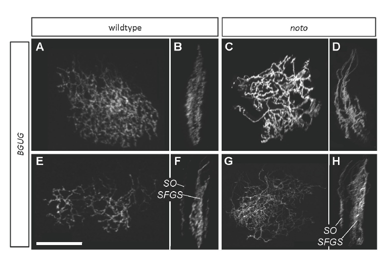
Fig. s3
BGUG+ ganflion cell (GC) axons are disorganized in 5 dpf noto mutant tecta. Images are projections of confocal stacks collected from live transgenic larvae. (A,B) Several wild-type SFGS GC axons in top (A) and side (B) views. (C,D) A similar number of mutant SFGS axons. Crossovers between sublaminae are evident. (E,F) Top and side views of a large group of wild-type GC axons, including one innervating SO. SO and SFGS axons are clearly separated. (G,H) A similar number of mutant axons, including one innervating SO. SFGS disorganization is apparent, and a dimly labeled axon appears to cross between SO and SFGS. This was the only observed instance of an SO-SFGS crossover. Scale bar: 50 μm.