
|
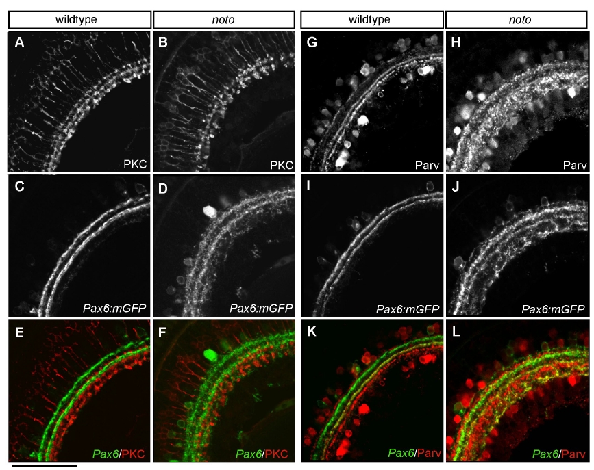
Fig. s2
Inner plexiform layer (IPL) sublamination defects in noto mutants become more severe by 7 dpf. (A,B) Protein kinase C (PKC)+ bipolar cells project terminals to three IPL sublaminae in wild type (A), whereas in the mutant (B) this targeting is coarse and the outermost sublamina remains sparse. (C,D) Pax6:mGFP+ neurites from the same sections shown in A and B, showing severely coarse targeting in the mutant. (E,F) Merged images showing the relative location of sublaminae. (G,H) Parvalbumin (Parv)+ amacrine cells project neurites to three IPL sublaminae in wild type (G), whereas in the mutant (H) this targeting has become very coarse. (I,J) Pax6:mGFP+ neurites from the same sections shown in G and H, showing severely coarse targeting in the mutant. (K,L) Merged images showing the relative location of sublaminae. Scale bar: 50 μm.