
|
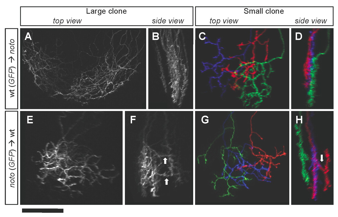
Fig. 6
noto mutant ganglion cell (GC) sublamination defects are retina-autonomous. (A-D) GC axons derived from wild-type zebrafish donors, innervating mutant tecta. (A,B) Top and side views of a large cluster of axons, including one SO and many SFGS axons, showing orderly sublamination. (C,D) Top and side views of individual GC axons, showing fine sublamination within SFGS. (E-H) GC axons derived from mutant donors, innervating wild-type tecta. (E,F) Top and side views of a large cluster of SFGS axons, showing disordered sublamination; arrows show laminar crossovers. (G,H) Top and side views of three SFGS axons. Red axon is multilaminar; arrow shows crossover. Scale bar: 50 μm.