
|
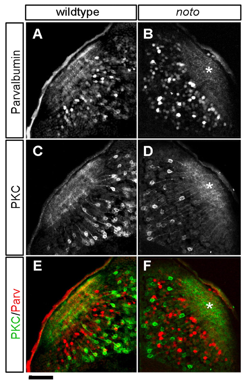
Fig. 3
noto mutant tectal neurons have unrefined neuropil projections at 5 dpf. Sections of wild-type (A,C,E) and mutant (B,D,F) zebrafish tectum showing tectal cell populations and their neuropil neurites. Disorganized neuropil in the mutant is marked with asterisks. (A,B) Wild-type Parvalbumin (Parv)+ tectal neurons project neurites to four laminae in the neuropil; this laminar organization is incomplete in the mutant. (C,D) Wild-type Protein kinase C (PKC)+ tectal neurons project neurites to three laminae in the neuropil; this laminar organization is lost in the mutant. (E,F) Merged images showing relative location of laminae. Scale bar: 50 μm.