
|
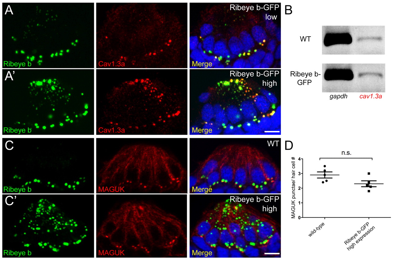
Fig. 10
Ectopic aggregates of overexpressed Ribeye colocalize with ectopic clusters of CaV1.3a channel in hair cells. Merged images include DAPI (blue). Scale bars: 3 μm. (A,A2) Ribeye b (green) and CaV1.3a (red) immunolabeling in cross-sections of anterior lateral line neuromasts in 6 dpf larvae. (B) RT-PCR analysis of cav1.3a transcripts in wild-type and transgenic larvae. gapdh was used as a loading control. (C,C2) Ribeye b (green) and MAGUK (red) immunofluorescent labeling in cross-sections of neuromasts. (D) The average number of MAGUK-positive punctae in wild-type and larvae stably expressing high levels of GFP-tagged Ribeye b. Error bars indicate s.e.m.