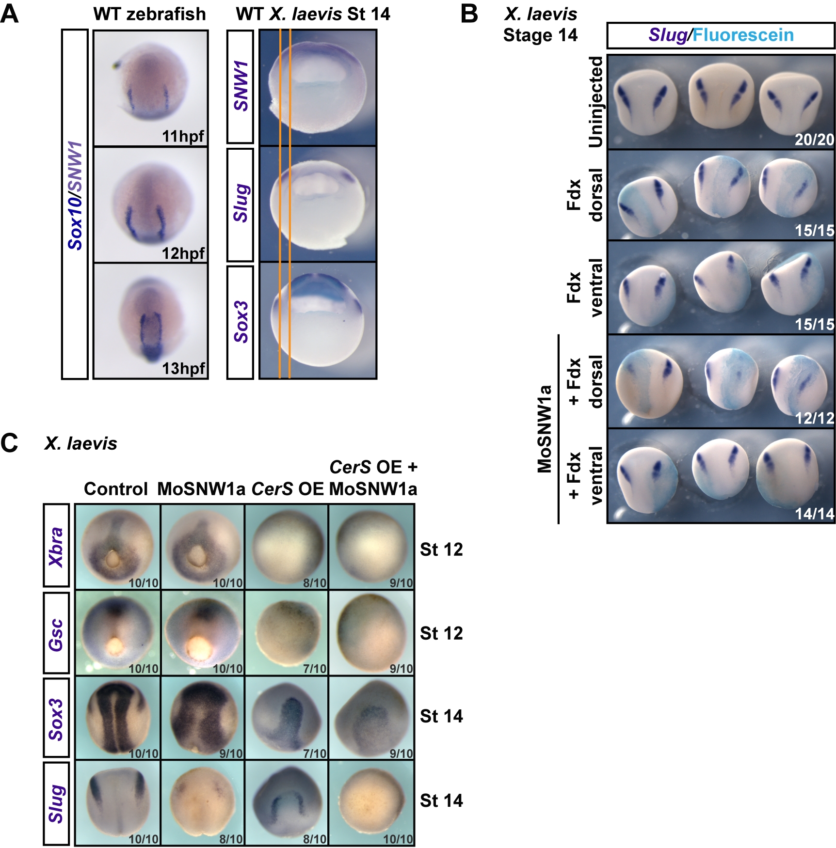
Fig. 3 Dorsally expressed SNW1 is required for neural crest specification, and this is independent of mesoderm.
(A) Left panels. Wild-type (WT) zebrafish embryos at the indicated times post-fertilization were doubly stained for the neural crest marker Sox10 (dark blue) and SNW1 (grey-purple), which is localized in the neural plate (see Figure 1F). Right panels. Wild-type stage 14 Xenopus embryos were stained for either SNW1, the neural crest marker Slug or the neural plate marker Sox3. The orange lines mark the edges of the neural crest staining to enable direct comparison of expression domains. (B) One dorsal or one ventral blastomere of four-cell Xenopus embryos was injected with Fdx with or without 10 ng of MoSNW1a, as indicated. At stage 14 the embryos were stained for Slug and fluorescein. (C) Xenopus embryos were either uninjected (control), injected with 20 ng of MoSNW1a at the one-cell stage, injected with 250 pg of CerS mRNA into all four blastomeres at the four-cell stage, or injected with MoSNW1a at the one-cell stage followed by CerS mRNA at the four-cell stage. WISH was carried out for the mesoderm markers Xbra and Gsc at stage 12, or the neural plate marker Sox3 and the neural crest marker Slug at stage 14. OE, overexpression. In all cases the number of embryos out of the total analyzed that showed the presented staining pattern is given.

