
|
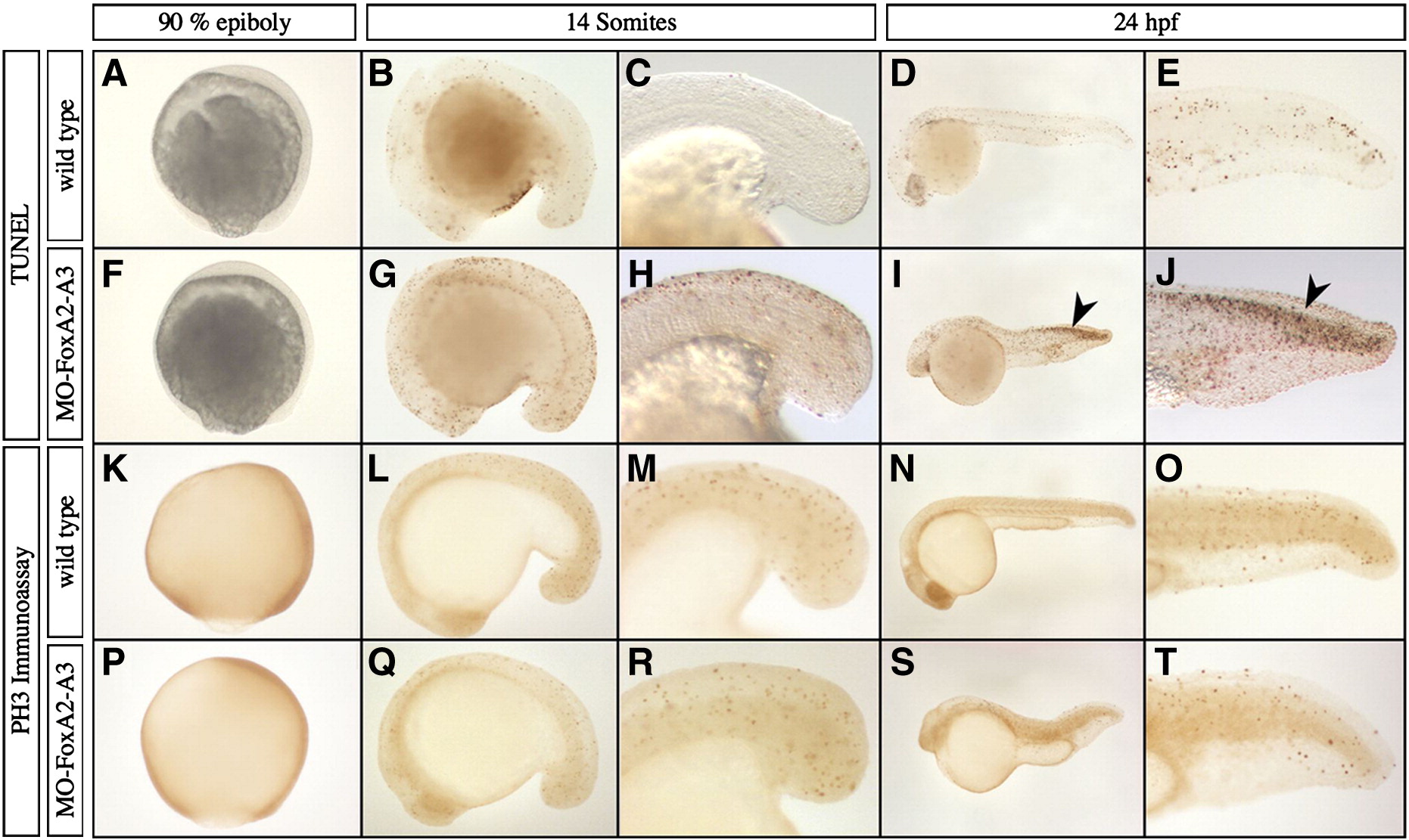
Fig. S3 Analysis of cell proliferation and apoptosis in FoxA2 and FoxA3 morphants. (A–J) Analysis of apoptosis by TUNEL assay at 90% epiboly, 14 somites and 24 hpf. The pattern of apoptotic cells in FoxA2–FoxA3 morphants is similar to wild-type embryos at every stage, except in the dorsal neural tube at 24 hpf, that shows strong apoptosis (arrowheads in I and J). (K–T) Analysis of cellular proliferation by phospho-histone H3 immunoassay at 90% epiboly, 14 somites and 24 hpf. FoxA2–FoxA3 morphants are similar to wild-type embryos at all stages. (A–T) Lateral views, anterior to the left (except in A, F, K, and P with animal pole up). (C, E, H, J, M, O, R, and T) Magnification of the tail region.
Reprinted from Developmental Biology, 350(2), Dal-Pra, S., Thisse, C., and Thisse, B., FoxA transcription factors are essential for the development of dorsal axial structures, 484-495, Copyright (2011) with permission from Elsevier. Full text @ Dev. Biol.