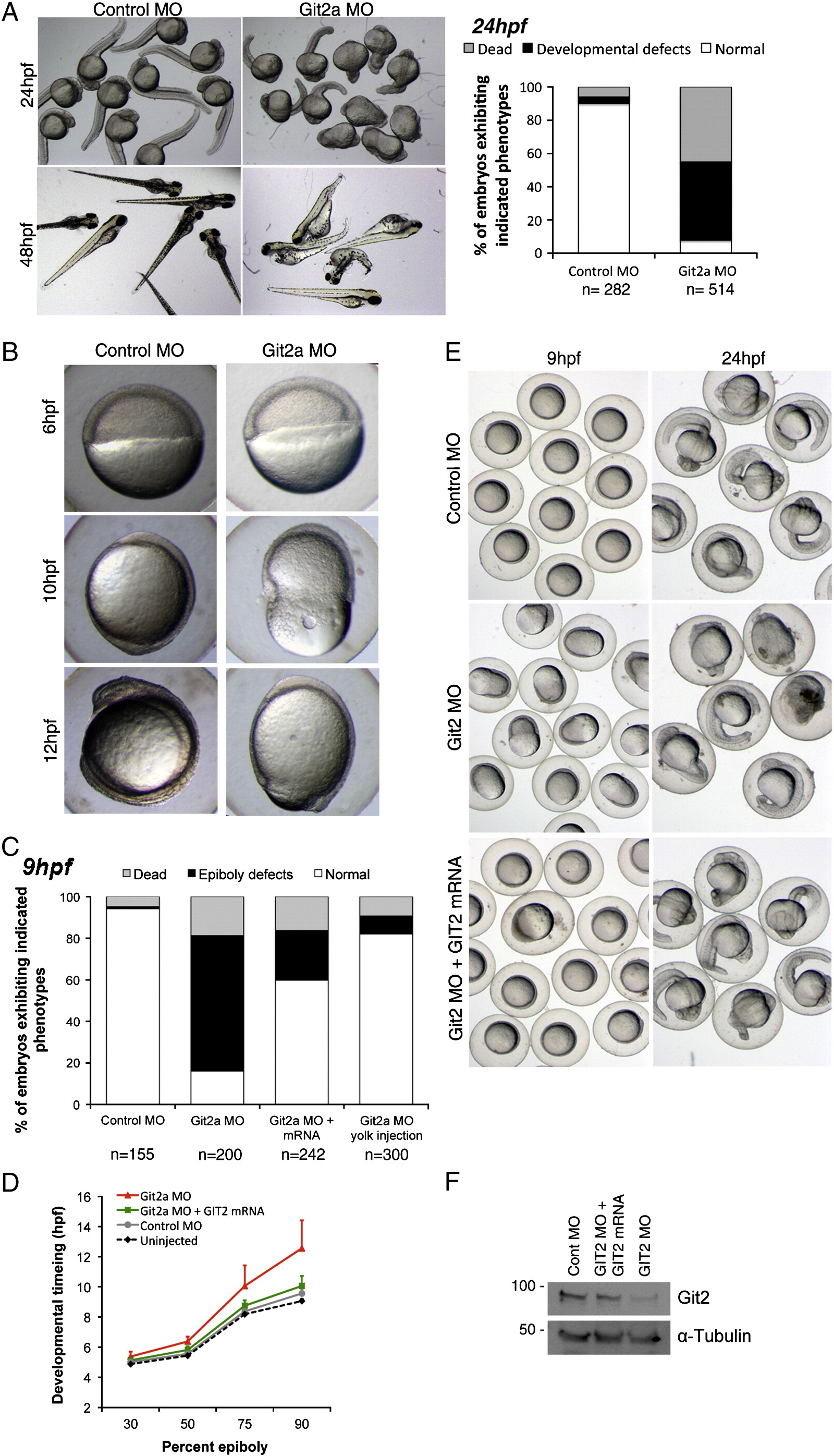
Fig. 2 Embryonic phenotypes following Git2a morpholino knockdown. (A) Live embryos injected with either control morpholino (MO) or git2a MO at 24 hpf and 48 hpf. git2a morphants exhibited variable defects including a curled tail, a short body axis and edema. The graph shows the quantification of phenotypes at 24 hpf from three independent experiments. (B) Representative images of control and git2a morphant embryos at 6, 10 and 12 hpf. A delay or arrest of epiboly was observed in git2a morphants. (C) The percentage of control and git2a MO embryos showing epiboly defects. Co-injection of chicken GIT2 mRNA partially rescued these defects. Injection of git2a MO into the yolk cell resulted in only minor epiboly defects. (D) Timing of epiboly progression in uninjected, control MO, git2a MO injected and chicken GIT2 mRNA rescued embryos. Data combined from at least four independent experiments. (E) Representative images of chicken GIT2 mRNA rescue phenotypes compared with control and git2a morphants at 9 and 24 hpf. (F) Western blot for Git2 protein expression in git2a morphants and embryos co-injected with git2a MO and chicken GIT2 mRNA.
Reprinted from Developmental Biology, 349(2), Yu, J.A., Foley, F.C., Amack, J.D., and Turner, C.E., The Cell Adhesion-associated Protein Git2 Regulates Morphogenetic Movements during Zebrafish Embryonic Development, 225-237, Copyright (2011) with permission from Elsevier. Full text @ Dev. Biol.

