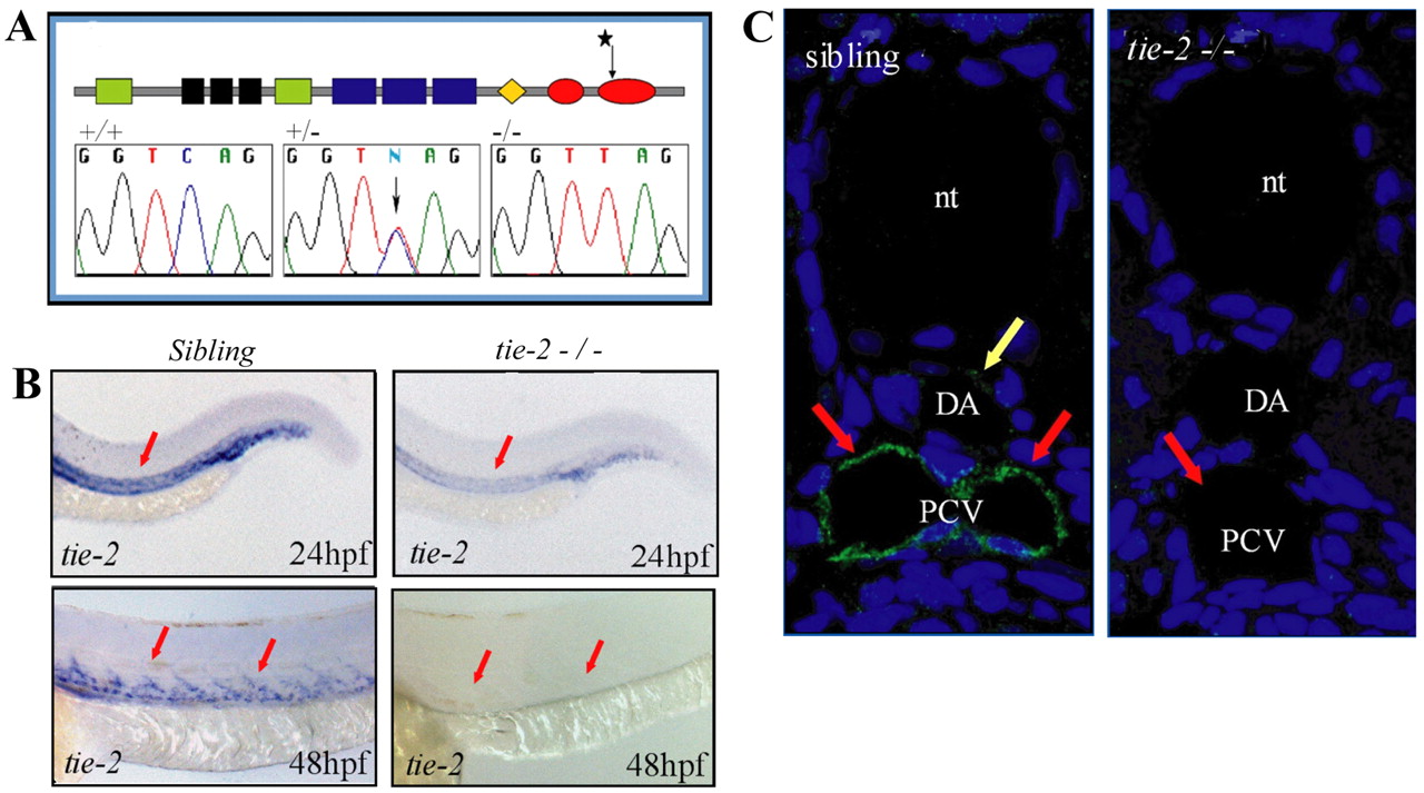
Fig. 1 A mutation in the kinase domain of tie-2 leads to loss of Tie-2 protein. (A) Schematic of the Tie-2 protein domains. Green, black and blue boxes represent the Ig-like, EGF-like and FN-III protein domains, respectively. The orange diamond represents the transmembrane region and the two red ovals represent the two kinase domains. Sequence chromatograms show the tie-2hu1667 allele to encode a stop codon within the second kinase domain. Sequence of the tie-2 hu1667 allele: CAG (Q982) is changed into a TAG (STOP) codon (mutation indicated by a star). (B) At 24 hpf, tie-2 mRNA expression levels are significantly reduced in tie-2 mutants compared with wild-type siblings (red arrows). tie-2 mRNA expression is absent at 48 hpf in mutants, whereas it is readily detectable in wild-type siblings (red arrows). (C) In wild-type fish, Tie-2 protein is expressed in the PCV at 48 hpf. Tie-2 protein expression is completely undetectable in tie-2 mutants. Yellow arrow shows Tie-2 protein expression in the dorsal aorta (DA); red arrows show Tie-2 protein expression in the PCV. nt, notochord.
Image
Figure Caption
Figure Data
Acknowledgments
This image is the copyrighted work of the attributed author or publisher, and
ZFIN has permission only to display this image to its users.
Additional permissions should be obtained from the applicable author or publisher of the image.
Full text @ Dis. Model. Mech.

