
|
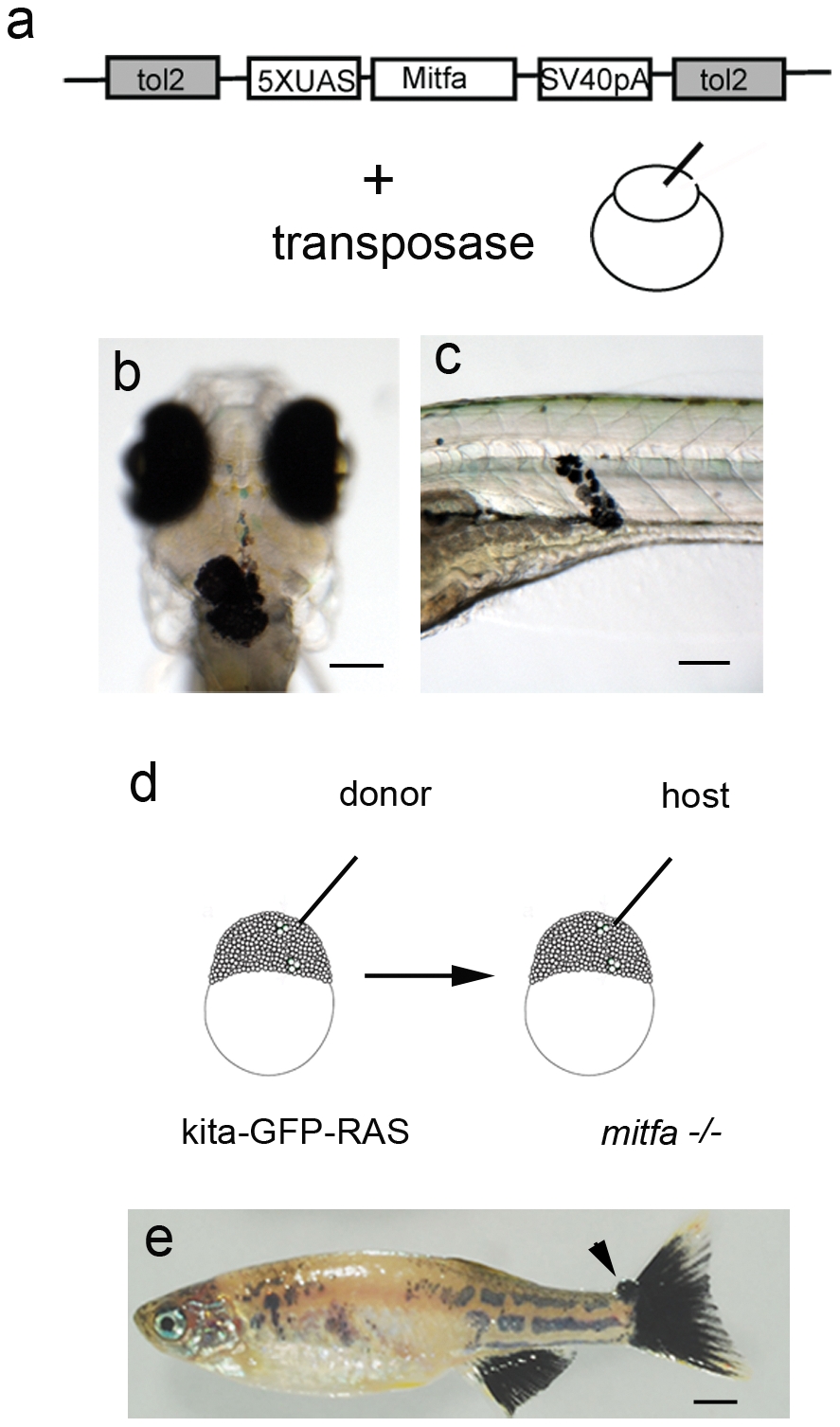
Fig. 6 Melanoma development is dependent on mitfa expression.
a) Schematic diagram of the construct and injection strategy used to re-express mitfa in mitfa-/- (nacre); kita-GFP-RAS zebrafish. Transformed melanocytes in the head of a 30hpf (b) and in the trunk of 5dpf (c) mitfa-/- (nacre); kita-GFP-RAS injected larvae. d) Schematic representation of the transplantion strategy, where cells were transplanted from kita-GFP-RAS embryos into mitfa-/- (nacre) embryos at approximately 1000 cell stage. e) Picture of 4 month-old trasplanted mitfa-/- (nacre) fish displaying a patchy recovered pigment pattern and a small tumor at the level of the caudal fin (arrow). Calibration bars = 100 μm for b,c; 2 mm for e.