
|
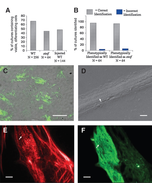
Fig. 7 Microinjection of embryos with antisense morpholino-oligonucleotides or gene expression constructs followed by seZEB culture. The percentage of cultures with viable, differentiating cells was measured for genotyped WT, steif, and injected embryos (A). In the reverse experiment, phenotypic identification of steif mutants and WT embryos was confirmed by dCAPS genotyping, demonstrating nearly 100% accuracy (B). Only a few cultures identified phenotypically as derived from WT/heterozygous or mutant embryos were found to be otherwise. (C) Zebrafish embryos were injected at the 2- to 4-cell stage with unc45b-GFP expression constructs. GFP expression was detectable in bundles of differentiating cells as early as 2 days of culture (scale bar = 0.5 mm). (D) Blastomeres from embryos injected with p53 control morpholinos were able to undergo attachment and differentiation in culture, resulting in the formation of bundled myocytes with visible striations (arrow, scale bar = 0.05 mm). (E, F) Embryos injected with a morpholino against unc45b were costained with Alexa 568 phalloidin (E, red) and F59 anti-myosin antibody detected by Alexa 488 anti-mouse (F, green). The punctate pattern of actin and myosin expression seen in steif mutant cultures was reproduced in nearly all cultures from unc45b morpholino-oligonucleotide-injected embryos (arrows). Scale bars (E, F) = 0.1mm.