
|
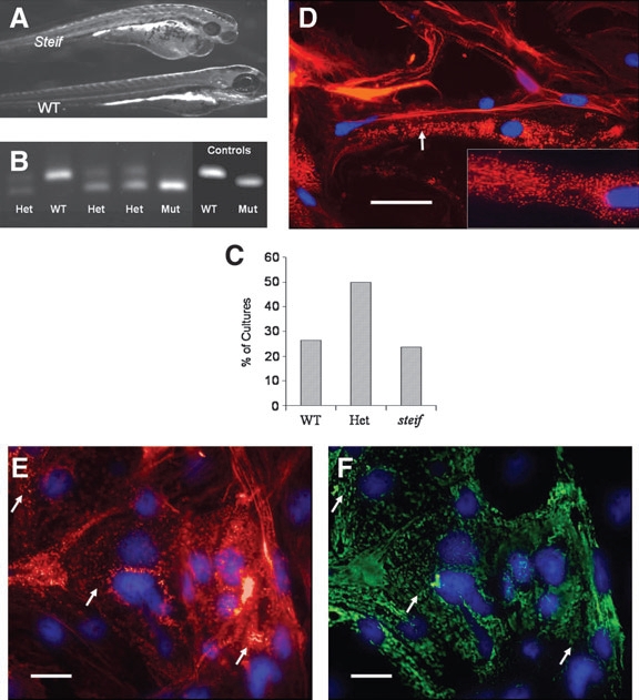
Fig. 6 Marker expression and genotyping of seZEB cultures from unc45b mutant embryos. Single- embryo cultures of ZEB cells were established from embryos obtained by crossing steif mutant heterozygote parents. (A) Phenotype of 3-day-old steif mutant embryo as compared to WT (bottom), demonstrating heart edema and reduced birefringence of tail muscle. Cell cultures from mutant embryos were identified by dCAPS genotyping of culture media (B), compared to control DNA amplified from phenotypically identified mutant embryos or WT adults. steif alleles detected in cultured embryos gave the expected 1:2:1 ratio of homozygous WT, heterozygous, and homozygous mutants (C, n = 88). Genotype-confirmed mutant seZEB cultures were stained for actin with Alexa 568 phalloidin (D) to observe sarcomere banding patterns (red), with DAPI as a nuclear counterstain (blue). Inset shows higher magnification. Mutant cultures demonstrated deficiencies in sarcomere organization and punctate expression of actin (arrow in D). (E, F) Costaining of actin (red) with the F59 anti-myosin antibody detected by Alexa 488 anti-mouse (green) demonstrated colocalization of disorganized, punctate actin and myosin expression (arrows). Scale bars in all panels = 0.1mm.