
|
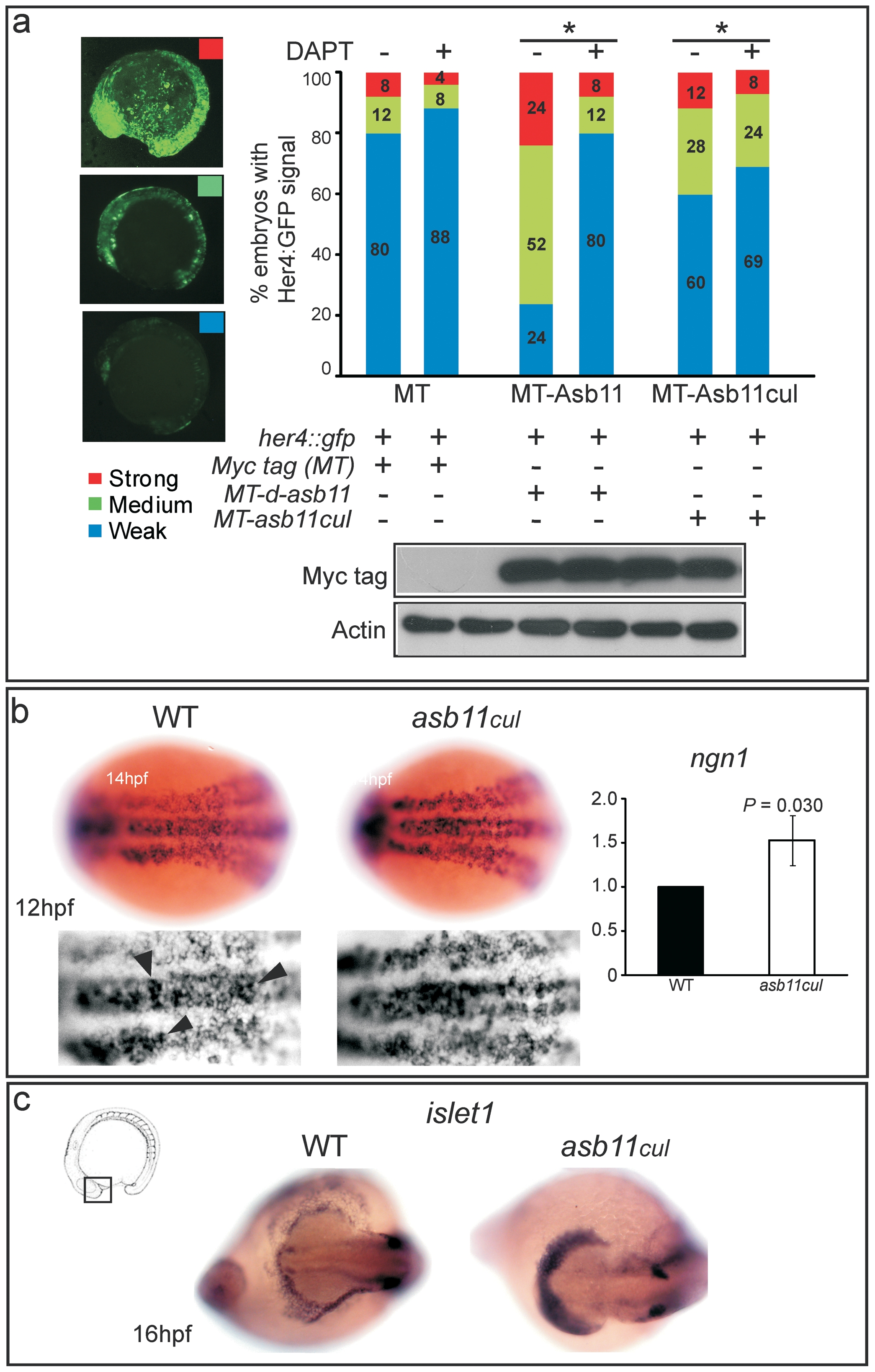
Fig. 5 her4::gfp transactivation and premature differentiation of neural cells in asb11cul.
(A), the her4::gfp reporter was co-injected with myc-tag (MT) mRNA as a control, myc-tagged d-asb11 full length (MT-Asb11) or myc-tagged asb11cul (MT-Asb11cul) mRNA in zebrafish embryos. Injected embryos were treated with (+) (n = 25) or without (-) (n = 25) DAPT, from 1.5 hpf. At 14 hpf, embryos were analyzed for her4 transactivation based on the intensity of the GFP signal. Positive embryos were counted and percentages of embryos presenting weak (blue), medium (green) or strong (red) signal were given. (B), Wild type (left panel) and mutant (middle panel) embryos at 12 hpf were analyzed for WISH using probe against ngn1. (right) Graph quantifies expression of ngn1 using qPCR. (C) Wild type (left panel) and mutant (right panel) polster of embryos at 16 hpf were analyzed for WISH using probe against islet1.