
|
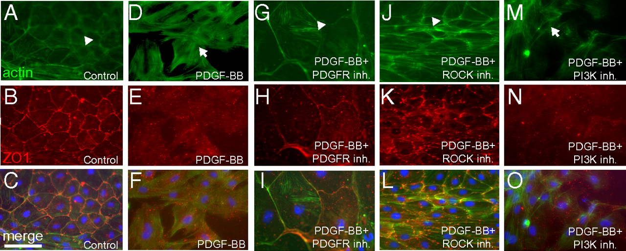
Fig. 4 PDGF signaling is sufficient to induce stress fibers and loss of cell-cell contacts in vitro. Sham-operated hearts were cultured in the presence of 0.5% serum for 3 d and then treated with 0.5% BSA as a control (n = 12 hearts) (A–C), PDGF-BB (n = 4 hearts) (D–F), PDGF-BB and PDGFR inhibitor (Inh.; 2 μM, n = 4 hearts) (G–I), PDGF-BB and ROCK inhibitor (2 μM, n = 4 hearts) (J–L), or PDGF-BB and PI3K inhibitor (2 μM, n = 4 hearts) (M–O) for 1 d (i.e., from day 3 to day 4 in culture) (actin, green; DAPI: blue; ZO1, red). Actin localized to the subcortical region is marked by arrowheads, and stress fibers are marked by arrows. Each individual explant was imaged in at least three different fields of view, and a representative image is shown. (Scale bar = 50 μm.)