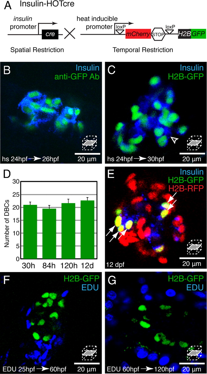
Fig. 3
Dorsal bud derived β-cells (DBCs) are quiescent. (A) Insulin-HOTcre, Heat-inducible Overexpression of Transgenes in an insulin: Cre restricted domain. Expression of Cre in β-cells excises the mCherrySTOP cassette and permits heat-inducible expression of H2B-GFP. (B–G) Insulin-HOTcre embryos were heat-shocked at 24 hpf. (B and C) Confocal sections through an islet stained for Insulin and GFP at 26 hpf (B) or Insulin at 30 hpf (C). (D) Quantification of H2B-GFP positive cells per embryo after 24 hpf heat-shock from 30 hpf to 12 dpf, mean ± SEM (Table S1). (E) Insulin-HOTcre embryos were injected with H2B-RFP mRNA at the one-cell stage, and heat-shocked at 24 hpf. Confocal section of the principal islet stained for Insulin at 12 dpf. The mCherry heat-shock induction marker is degraded by 12 dpf (See Fig. S2A). (F and G) Embryos were labeled by pericardial EDU injection at 25 (F) or 60 (G) hpf. Confocal sections through islets at 60 (F) or 120 (G) hpf. (B) All Insulin-positive cells coexpress H2B-GFP at 26 hpf. (C) β-cells that differentiated after the heat-shock at 24 hpf express Insulin but do not express H2B-GFP (arrowhead). (D and E) The number of H2B-GFP positive cells after a 24 hpf heat-shock does not significantly change during development (D). Furthermore, all H2B-GFP-positive cells labeled after heat-shock at 24 hpf, also retain the H2B-RFP label (arrows, E). (F and G) Many cells within the pancreas divide during embryonic and larval development (blue nuclei). However, β-cells labeled by Insulin-HOTcre induction at 24 hpf did not exhibit EDU incorporation (n = 5 islets for each condition).

