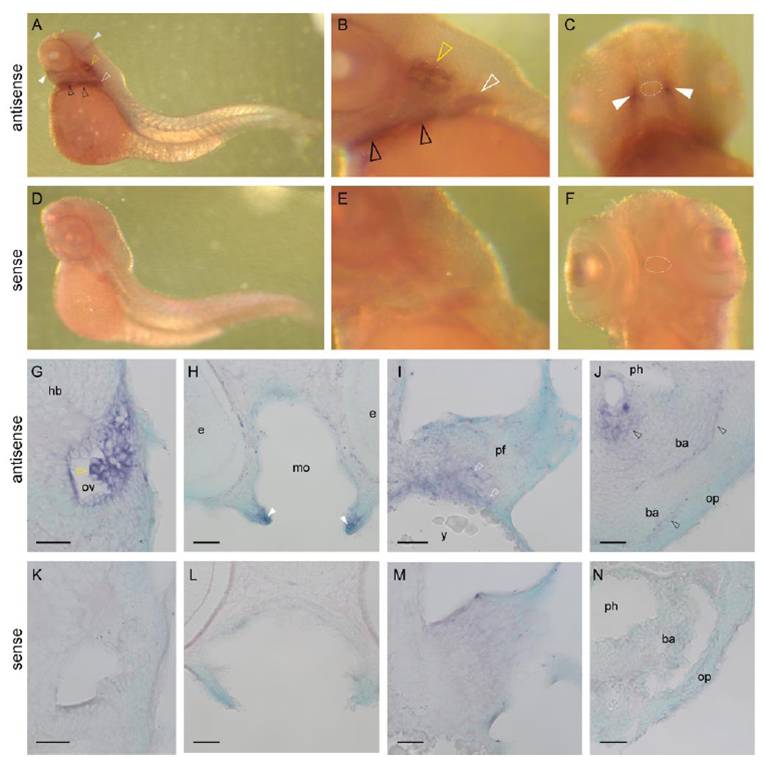
Fig. S3 Expression pattern of gnav1 in zebrafish larvae: whole-mount in situ hybridization of gnav1 probe (A–C and G–J) and negative control (sense probe, D–F and K–N) with 3 dpf zebrafish larvae. Embryos were grown in 0.0045% of phenylthiourea (Sigma) from 12 h postfertilization until used to prevent pigmentation. Probes were synthesized with T3 RNA polymerase (Roche) by following manufacturer’s instructions. Templates for in vitro transcription were amplified using primers listed in Table S3. Probes were hybridized at 65 °C overnight. Specific probe hybridization was detected with anti-DIG antibody conjugated with alkaline phosphatase (1:5,000; Roche), using Nitroblue tetrazolium chloride/5-bromo-4-chloro-3-indolyl-phosphate (Roche) as substrates. Stained larvae were observed and photographed with a Nikon SMZ-U binocular and an attached Nikon CoolPix 950 digital camera. Stained larvae were cryo-sectioned (8 μm), counterstained in 0.001% methyl green, mounted with VectaMount (Vector), and documented on a Zeiss Axioplan microscope and an attached AxioCamMRc5. (A and D) Lateral view of whole larvae. (B and E) lateral view around the developing inner ear. (C and F) Ventral view of head region. Dotted circle, mouth. (G–N) Cross-sections of stained larvae at the levels of developing inner ear (G and K), mouth (H and L), pectoral fin (I and M), and branchial arches (J and N). Dorsal is to the top. White and gray arrowheads indicate the cell clusters near the lip and midbrain–hindbrain boundary, respectively. White, yellow, and black arrowheads point to labeled cells within pectoral fin (pf), otic vesicle (ov), and branchial arches (ba), respectively. e, eye; hb, hindbrain; mo, mouth cavity; op, operculum; ph, pharynx; y, yolk. (Scale bars: 50 μm.)
Image
Figure Caption
Acknowledgments
This image is the copyrighted work of the attributed author or publisher, and
ZFIN has permission only to display this image to its users.
Additional permissions should be obtained from the applicable author or publisher of the image.
Full text @ Proc. Natl. Acad. Sci. USA

