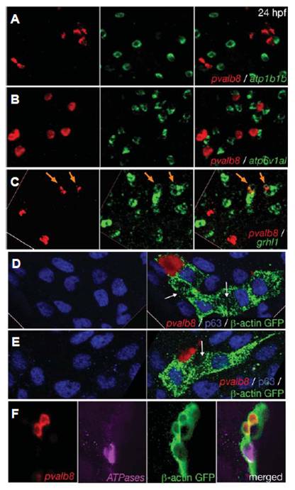
Fig. 4 pvalb8-expressing cells are distinct from NaR and HR ionocytes, but also display transient grhl1 expression and derive from the common pool of epidermal progenitors. All panels show in situ hybridisations and/or immunostainings with the probes and antibodies indicated in lower right corners. (A,B) Double fluorescent in situ hybridisations of wild-type embryos at 24 hpf, revealing that pvalb8- positive cells lack expression of atp1b1b (A) and atp6v1al (B). (C) Double fluorescent in situ hybridisations of wild-type embryos at 24 hpf, revealing co-expression of grhl1 and pvalb8 in cells indicated by orange arrows. The left and middle pictures of each panel show the single channels of the merged image shown in the right picture. (D-F) Clones of epidermal cells of wild-type embryos at 24 hpf, after homotopic and homochronic transplantation of single GFP-transgenic cells into the ventral ectoderm at 6 hpf (shield stage). The left pictures of (D,E) show an anti-p63 immunostaining of keratinocytes (in blue), the right pictures show an overlay of the p63 immunostaining with an in situ hybridisation for pvalb8 transcripts (in red) and an anti-GFP immunostaining for the descendants of the transplanted cell (in green). The white arrows point to p63, pvalb8- double negative cells in the clone, which might represent an NaR or HR ionocyte. (F) From left to right: in situ hybridization for pvalb8 transcripts (in red), in situ hybridization for atp1b1b and atp6v1al transcripts (in pink), anti-GFP immunostaining for the descendants of the transplanted cell (in green), merged image.
Image
Figure Caption
Figure Data
Acknowledgments
This image is the copyrighted work of the attributed author or publisher, and
ZFIN has permission only to display this image to its users.
Additional permissions should be obtained from the applicable author or publisher of the image.
Full text @ Int. J. Dev. Biol.

