Image
Figure Caption
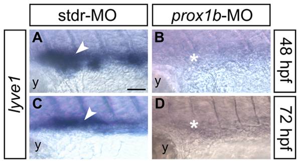
Fig. 4 The expression of the lymphatic molecular marker lyve1 is dramatically reduced in prox1b morphants.
lyve1 riboprobe labels developing lymphatic endothelial cells in 48 (A,B), and 72 (C,D) hpf embryos (all lateral views, anterior to the left; the approximate region of the embryo trunk imaged in all panels corresponds to the green box in Figure 3M). In comparison to control embryos injected with the stdr-MO (A,C), that display a normal expression of lyve1 (white arrowheads), the MO directed against prox1b (B,D) results in the absence of lyve1 signal (white asterisks). y, yolk. Scale bar represents 40 μm.
Figure Data
Acknowledgments
This image is the copyrighted work of the attributed author or publisher, and
ZFIN has permission only to display this image to its users.
Additional permissions should be obtained from the applicable author or publisher of the image.
Full text @ PLoS One

