Image
Figure Caption
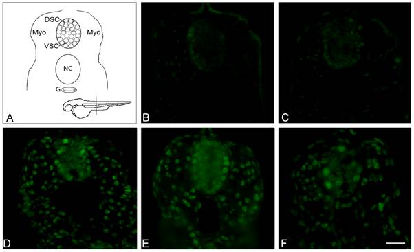
Fig. 5 TDP-43 localization in zebrafish embryos.
Immunofluorescent staining of endogenous zebrafish TDP-43 and overexpressed human TDP-43 was performed in transversely sectioned 30 hpf embryos in order to allow imaging of the spinal cord (shown in schematic diagram, A). TDP-43 localization was nuclear in all embryos examined (B: non injected, C: PGRN MO injected, D: Wt TDP-43 injected, E: Mt TDP-43 (A315T) injected, and F: co-expressing Mt TDP-43 and PGRN). The scale bar indicates a distance of 25 μm. Abbreviations: DSC, Dorsal spinal cord; VSC, Ventral spinal cord; Myo, myotomes; NC, notochord; G, gut.
Figure Data
Acknowledgments
This image is the copyrighted work of the attributed author or publisher, and
ZFIN has permission only to display this image to its users.
Additional permissions should be obtained from the applicable author or publisher of the image.
Full text @ PLoS One

