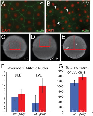
Fig. 5 poky mutants fail to down regulate cell division in the EVL. Embryos were stained with phalloidin (green) to reveal actin localization at cell boundaries and DAPI (red) to reveal nuclear morphology. Dividing cells were identified by their nuclear morphology (arrowheads, A,B). Multinucleate EVL cells were observed in wild type and mutant embryos (arrow, B). Total number of EVL cells was calculated by determining the number of cells per unit area as C and D and then calculating the total EVL surface area by calculating the radius and height as in E. (F) Percent mitotic nuclei. (G) Total number of EVL cells
Reprinted from Developmental Biology, 346(2), Fukazawa, C., Santiago, C., Park, K.M., Deery, W.J., Canny, S.G., Holterhoff, C.K., and Wagner, D.S., poky/chuk/ikk1 is required for differentiation of the zebrafish embryonic epidermis, 272-283, Copyright (2010) with permission from Elsevier. Full text @ Dev. Biol.

