
|
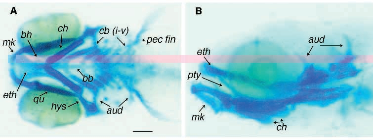
Fig. 1 Pharyngeal skeleton of a day- 5 zebrafish larva stained for cartilage with Alcian blue. (A) Ventral view. Individual cartilaginous elements of the pharyngeal skeleton are identifiable. The two trabeculae fuse medially to form the ethmoid plate (eth). First arch derivatives are Meckel’s cartilage (mk) and quadrate (qu). Second arch derivatives are basihyal (bh), ceratohyal (ch), hyosymplectic (hys). Gill arch derivatives are basibranchials (bb) and ceratobranchials I-V (cb i-v). Also labeled are cartilages of the pectoral fins (pec fin). (B) Lateral view reveals the pterygoid process of the quadrate (pty) and the auditory capsule (aud). Scale bar is 100 μm.