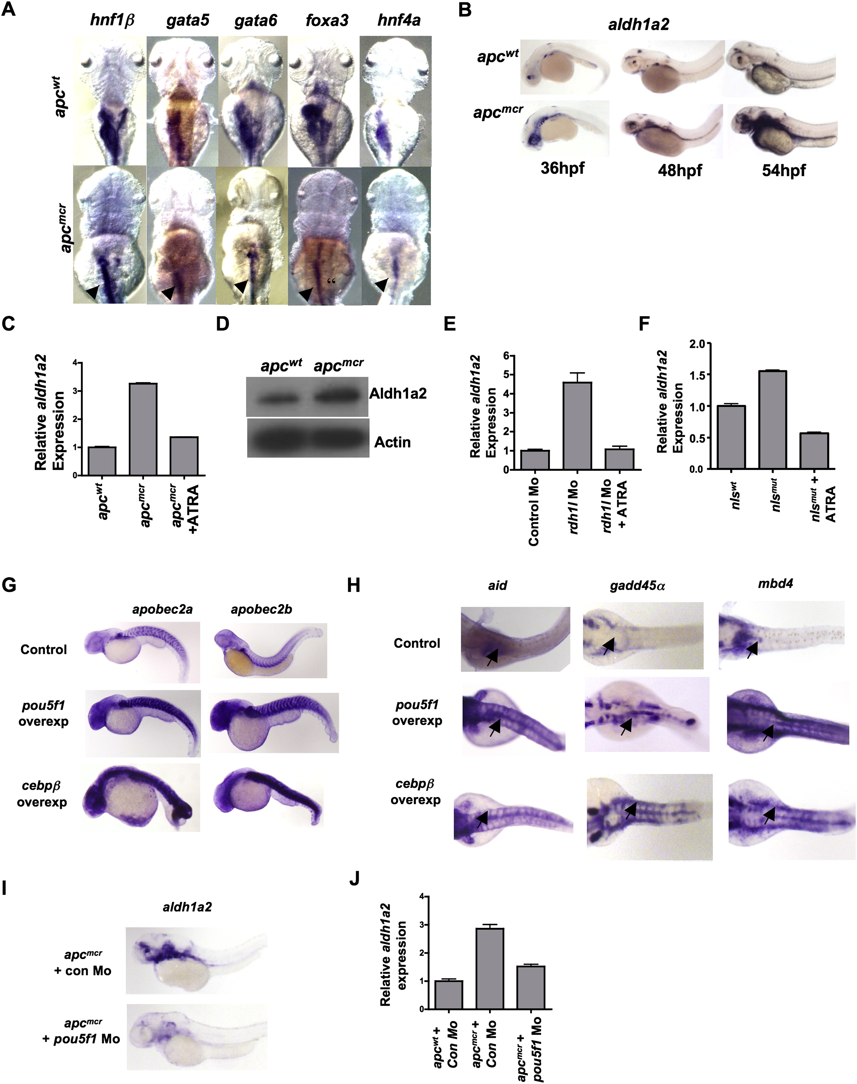
Fig. S3
Retinoic Acid and Pou5f1 Regulate aldh1a2 and Demethylase Gene Expression in apc Mutant Zebrafish Embryos, Related to Figure 3
(A) Whole mount in situ staining for hnf1β, gata5, gata6, foxa3 and hnf4α in apcmcr and apcwt embryos at 72hpf.
(B) Whole mount in situ staining for aldh1a2 in apcmcr and apcwt embryos at 36hpf, 48hpf and 56hpf.
(C) Graph showing fold change obtained from RT-PCR for aldh1a2 expression in apcmcr and apcwt embryos treated with DMSO or ATRA.
(D) Immunoblot analysis for Aldh1a2 in apcwt and apcmcr zebrafish embryos at 72hpf. Actin is used as a loading control.
(E) Graph showing fold change obtained from RT-PCR for aldh1a2 expression in control morphants and rdh1l morphants treated with DMSO or ATRA.
(F) Graph showing fold change obtained from RT-PCR for aldh1a2 expression in nls (neckless) wild-type or nls mutant embryos treated with DMSO or ATRA.
(G and H) Whole mount in situ staining for apobec2a and apobec2b (G) and aid, mbd4 and gadd45a (H) in embryos injected with control, pou5f1 or cebpβ expressing plasmids. In panel H, black arrows show expression in the intestine.
(I) Whole mount in situ staining for aldh1a2 in apcmcr and apcwt (at 72hpf) injected with control Mo or pou5f1 Mo (80pg).
(J) Graph showing fold change obtained from RT-PCR for aldh1a2 expression in apcmcr and apcwt embryos injected with control morpholino or pou5f1 morpholino. y axis values are fold changes in expression of indicated genes normalized first to 28S levels and then to mRNA/28S ratio from control samples, valued as 1.
Error bars are +/- SD.
Reprinted from Cell, 142(6), Rai, K., Sarkar, S., Broadbent, T.J., Voas, M., Grossmann, K.F., Nadauld, L.D., Dehghanizadeh, S., Hagos, F.T., Li, Y., Toth, R.K., Chidester, S., Bahr, T.M., Johnson, W.E., Sklow, B., Burt, R., Cairns, B.R., and Jones, D.A., DNA demethylase activity maintains intestinal cells in an undifferentiated state following loss of APC, 930-942, Copyright (2010) with permission from Elsevier. Full text @ Cell

