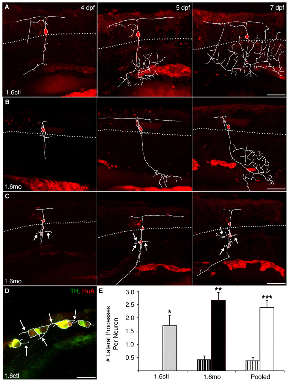
Fig. 4 Migratory DRG neurons sprout novel lateral processes. DRG neurons were electroporated with Alexa568 at 4 dpf and followed to 7 dpf. In panels A-C, dye-labeled DRG processes were traced in white from individual slices and overlaid onto the projected images. The dotted gray line represents the ventral boundary of the spinal cord, where DRG are typically found. (A) In a control embryo, a DRG neuron extends a single process ventrally at 4 dpf. At subsequent times, the process begins to branch once it reaches more ventral positions. (B) In a nav1.6 morphant embryo, a correctly positioned DRG neuron also extends a ventral process that branches. (C) By contrast, ectopic DRG neurons of morphant embryos sprout processes (white arrows) that emanate from the soma and extend laterally. (D) In an 11 dpf control embryo, laterally projecting processes emerge from the soma of TH+/HuA+ sympathetic neurons (white arrows). (E) Normally positioned DRG neurons (striped bars), in either control (grey) or morphant (black) embryos, sprout few or no laterally projecting processes from their soma. By contrast, migratory DRG neurons (solid bars) display many more laterally projecting processes (*P<0.05 versus normally-positioned 1.6ctl; **P<0.001 versus normally-positioned 1.6mo; ***P<0.001 versus normally positioned pooled cells; Kruskal-Wallis nonparametric ANOVA). White bars represent pooled morphant and control cells. Scale bars: 60 μm in A-C; 20 μm in D.
Image
Figure Caption
Figure Data
Acknowledgments
This image is the copyrighted work of the attributed author or publisher, and
ZFIN has permission only to display this image to its users.
Additional permissions should be obtained from the applicable author or publisher of the image.
Full text @ Development

