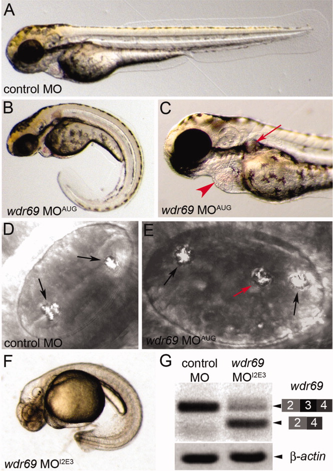
Fig. 2 wdr69 morpholino oligonucleotides (MO) knockdown embryos develop phenotypes associated with defects in ciliary motility. A-C: Live embryos at 2 days postfertilization (dpf). Control MO injected embryos exhibited normal morphology (A), whereas wdr69 MOAUG morphant embryos developed phenotypes associated with cilia defects, including a curled tail (B), kidney cysts (arrow in C) and pericardial edema (arrowhead in C). D,E: Otoliths in otic vesicles at 2 dpf. Control MO injected embryos developed two correctly positioned otoliths (arrows in D). wdr69 MOAUG morphants often developed a third ectopically positioned otolith (red arrow in E). F:wdr69 MOI2E3 morphants developed the same phenotypes as wdr69 MOAUG morphants (this embryo was treated with PTU to inhibit melanin biosythesis for RNA in situ analysis). G: RT-PCR analysis shows wdr69 MOI2E3 causes mis-splicing of wdr69 transcripts. A normally spliced cDNA containing exons 2-4 was detected in control MO injected embryos. The level of this cDNA was reduced in wdr69 MOI2E3 morphants and a smaller fragment lacking exon 3 was observed in wdr69 MOI2E3 morphants. β-actin was amplified as a loading control.
Image
Figure Caption
Figure Data
Acknowledgments
This image is the copyrighted work of the attributed author or publisher, and
ZFIN has permission only to display this image to its users.
Additional permissions should be obtained from the applicable author or publisher of the image.
Full text @ Dev. Dyn.

