Image
Figure Caption
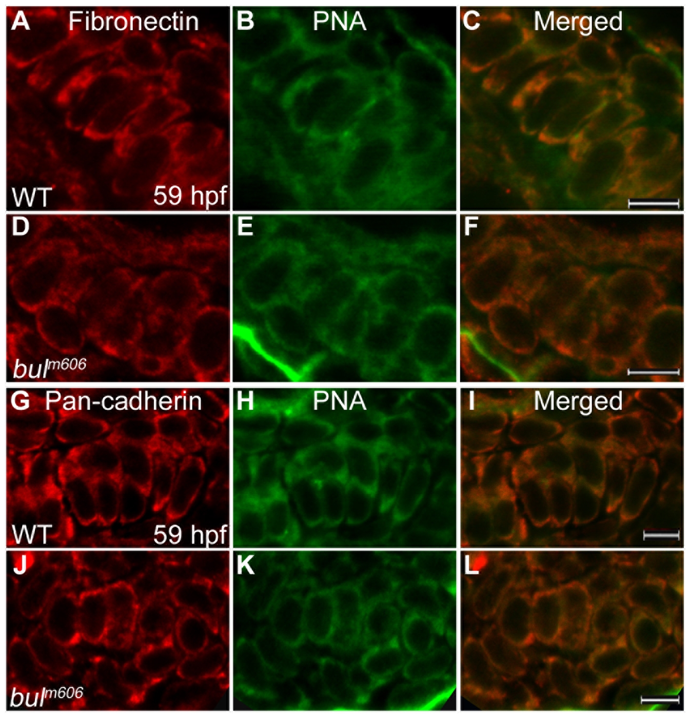
Fig. 5 Analysis of mesenchymal condensations in cartilage primordia of bulldog mutants.
(A–F) Mesenchymal condensations, stained with Peanut Agglutinin (PNA, green) show similar distribution of Fibronectin (red) in wild-type (A–C) and bulldog hyosymplectic cartilages (D–F). (G–L) Cadherin expression in chondrocytes (marked by a Pan-cadherin antibody in red) is indistinguishable between wild-type and bulldog ceratohyals as shown by PNA staining. The right panels represent merged images of the left and middle panels. Scale bars are 5 μM.
Figure Data
Acknowledgments
This image is the copyrighted work of the attributed author or publisher, and
ZFIN has permission only to display this image to its users.
Additional permissions should be obtained from the applicable author or publisher of the image.
Full text @ PLoS One

