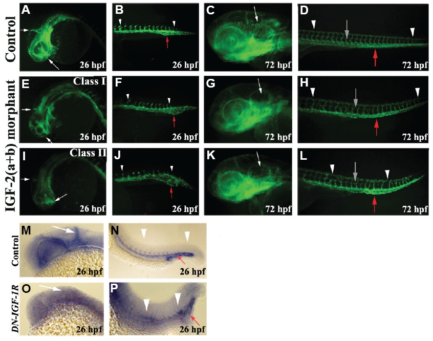
Fig. 5 Angiogenesis is compromised when IGF signalling is reduced. (AD) fli1:EGFP transgenic embryos injected with control morpholinos showing normal vascular development. (E-H) Class I IGF-2 morphant embryo. The intensity of GFP expressing cells in the head and eyes is reduced while the basic pattern of vasculature remains intact. The intermediate cell mass is mildly expanded and the intersomitic vessels are reduced. By 72 hpf, the parachordal vessel is incompletely formed. (I-L) Class II IGF-2 morphant embryo. The sprouting of vessels in the head and eyes are reduced. Intersomitic vessel sprouting is irregular and reduced, the intermediate cell mass is expanded and the parachordal vessel is disrupted. (M,N) Control embryo at 26 hpf showing normal expression of flk1 in the vasculature. (O,P) Angiogenesis is disrupted in DN-IGF-1R injected embryos as shown by flk1 expression (n=14/29). White arrows indicate the vasculature in the head and eyes. White arrowheads point to the intersomitic vessels, red arrow points to the intermediate cell mass and the grey arrow indicates the parachordal vessel. All embryos are shown in a lateral view.
Image
Figure Caption
Figure Data
Acknowledgments
This image is the copyrighted work of the attributed author or publisher, and
ZFIN has permission only to display this image to its users.
Additional permissions should be obtained from the applicable author or publisher of the image.
Full text @ Int. J. Dev. Biol.

