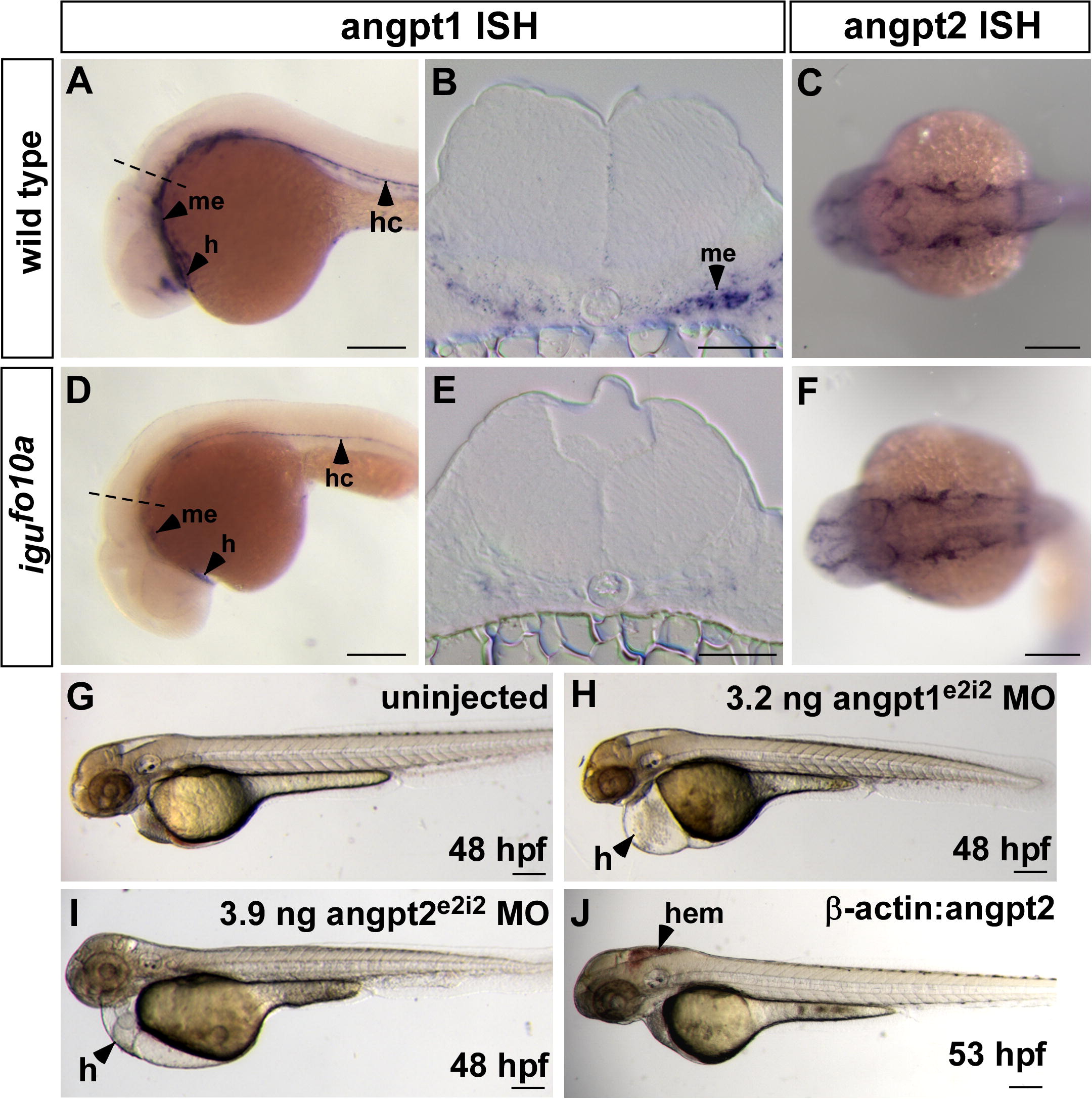
Fig. 4 Loss of Angiopoietin1–Tie signaling in igufo10a mutants results in hemorrhage. (A–F) (A and B) In wild type embryos at 28 hpf, angpt1 is expressed in the developing heart (h), hypochord (hc), and ventral mesenchyme (me) as shown in whole mount (A) and in cross-section (B). (D and E) In igufo10a mutants of the same age, angpt1 expression persists in the heart and hypochord, but is substantially down-regulated in ventral mesenchyme, as shown in whole mount (D) and in cross-section (E). The dashed line in (A) and (D) marks the level of the section in (B) and (E), respectively. (C and F) Expression of angpt2 is unchanged in igufo10a mutant embryos (F) compared to wild type sibs (C) at 28 hpf. (G–I) Injection of 3.2 ng of angpt1e2i2 MO (H) or 3.9 ng of angpt2e2i2 MO (I) into wild type embryos results in defective heart development, pericardial edema (H), and lack of circulation when compared to uninjected control embryos at 48 hpf (G). (J) Transgenic over-expression of angpt2 under the β-actin promoter results in hemorrhage (hem) in wild type embryos at 53 hpf. Scale bar is 500 μm in (A), (C), (D), and (F)–(J) and 50 μm in (B) and (E).
Reprinted from Mechanisms of Development, 127(3-4), Lamont, R.E., Vu, W., Carter, A.D., Serluca, F.C., MacRae, C.A., and Childs, S.J., Hedgehog signaling via angiopoietin1 is required for developmental vascular stability, 159-168, Copyright (2010) with permission from Elsevier. Full text @ Mech. Dev.

