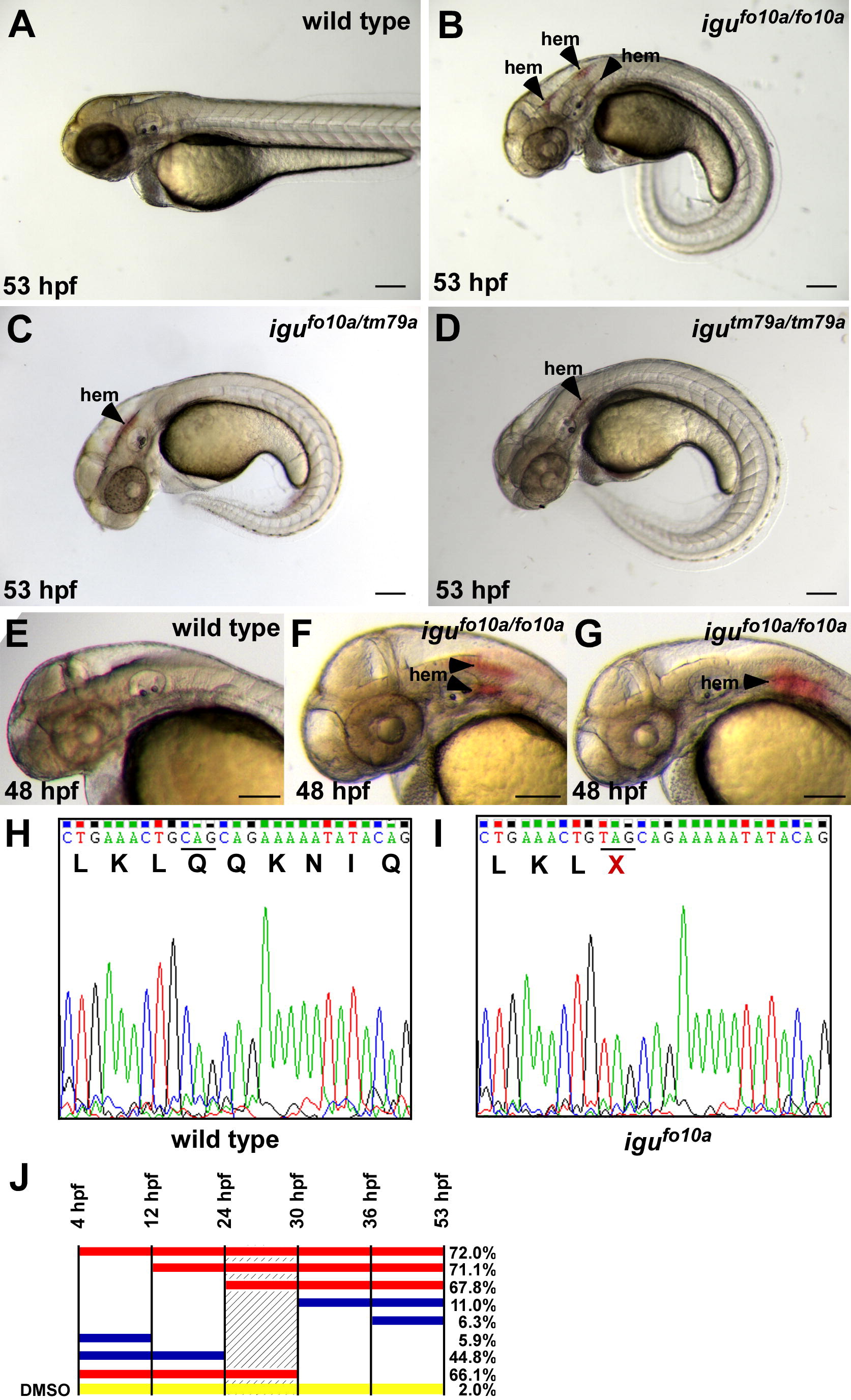
Fig. 1 Loss of hedgehog signaling leads to vascular stability defects. (A–D) igufo10a mutants present with a curled body axis and hemorrhage (hem) at 53 hpf (B), as compared to wild type siblings (A). igutm79a/igufo10a compound heterozygous embryos (C) and igutm79a mutant embryos (D) both present with hemorrhage at 53 hpf. (E–G) While wild type siblings do not show any signs of hemorrhage at 48 hpf (E), hemorrhages are present in the head of igufo10a mutants (F and G). (H and I) Electropherograms of dzip1 sequence from wild type (H) and igufo10a mutants (I) showing the presence of a Q339X mutation. (J) Hemorrhage rate after treatment with cyclopamine for developmental windows. Solid bars indicate the treatment interval with the percent of embryos displaying hemorrhage at 53 hpf given on the right side. Red bars indicate a treatment interval that results in maximal hemorrhage rates, while blue bars indicate intervals that produce minimal hemorrhage. The yellow bar indicates DMSO controls treated for the entire interval. The shaded area represents the most sensitive interval to cyclopamine treatment. Scale bar in (A)–(G) is 500 μm.
Reprinted from Mechanisms of Development, 127(3-4), Lamont, R.E., Vu, W., Carter, A.D., Serluca, F.C., MacRae, C.A., and Childs, S.J., Hedgehog signaling via angiopoietin1 is required for developmental vascular stability, 159-168, Copyright (2010) with permission from Elsevier. Full text @ Mech. Dev.

