
Image
|
Figure Caption
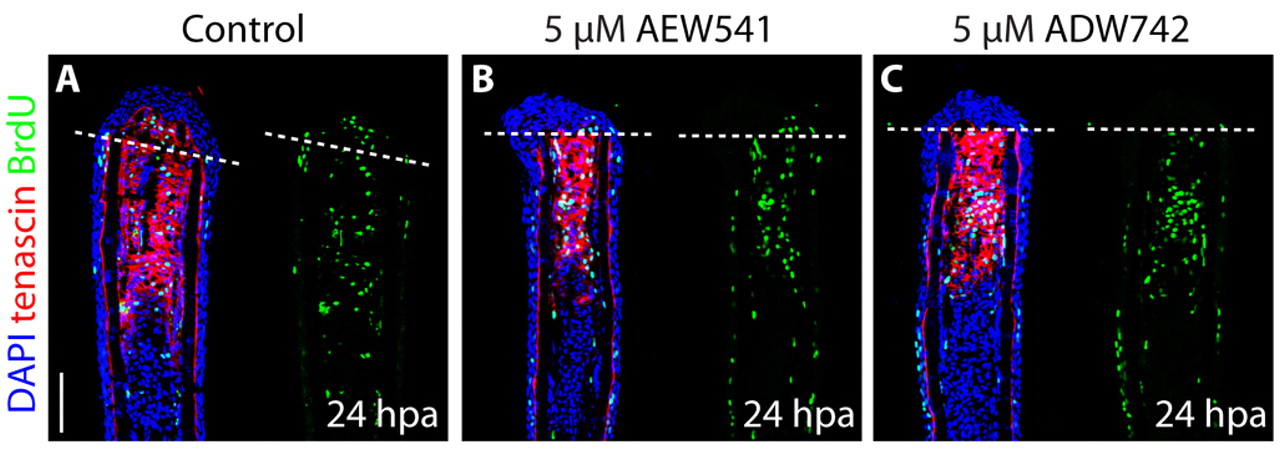
Fig. 7 Inhibition of IGF signaling does not affect cell proliferation and Tenascin C expression at the onset of blastema formation. (A-C) Fin regenerates at 24 hpa triply stained with BrdU antibody (green), Tenascin C antibody (red) and DAPI (blue). The dashed line demarcates the amputation plane. At this stage of regeneration, no difference in cell proliferation or Tenascin C expression is detected between the control (A) and Igf1r inhibitor-treated fins (B,C). Scale bar: 50 μm.
Figure Data
Acknowledgments
This image is the copyrighted work of the attributed author or publisher, and
ZFIN has permission only to display this image to its users.
Additional permissions should be obtained from the applicable author or publisher of the image.
Full text @ Development