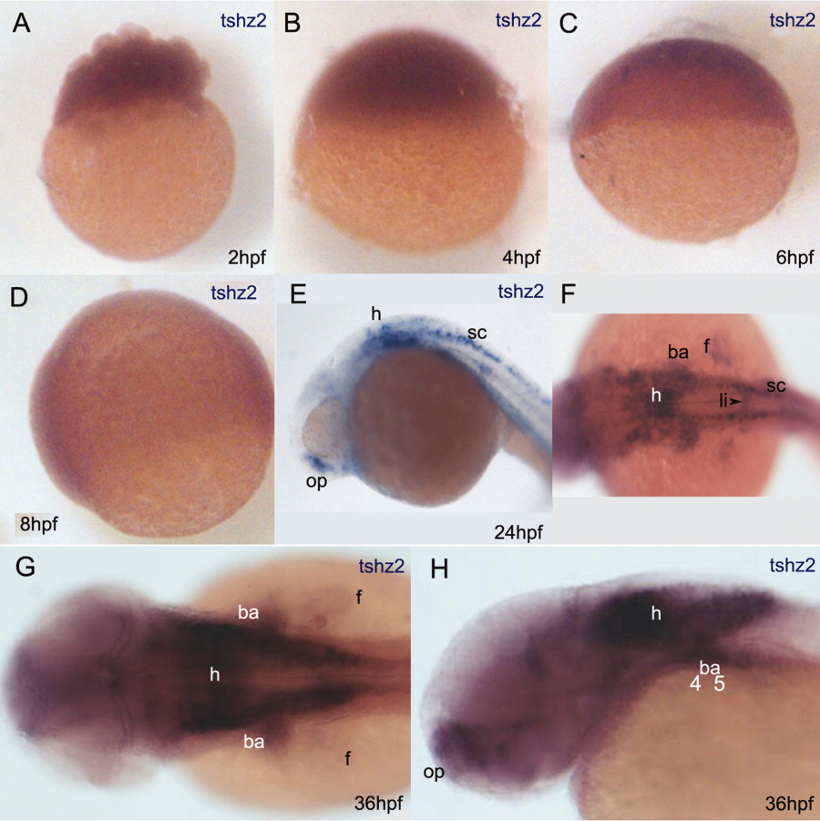
Fig. 2 Spatial and temporal expression pattern of tshz2 detected by RNA in situ hybridization. A-D: Embryos at 2 (A), 4 (B), 6 (C), and 8 (D) hours postfertilization (hpf). tshz2 mRNA is detected in early stages but signal fades by 8-10 hpf. Re-expression of tshz2 occurs around the prim-5 stage (24 hpf) (E,F). G,H: tshz2 expression pattern in a 36 hpf embryo. At 36 hpf, tshz2 is also expressed in the liver primordium (not shown). F,G: Dorsal views with anterior to the left. (E and H) Lateral views with dorsal up and anterior to the left. ba, branchial arches; f, pectoral fin; h, hindbrain; l, liver; op, olfactory placode; sc, spinal cord.
Image
Figure Caption
Figure Data
Acknowledgments
This image is the copyrighted work of the attributed author or publisher, and
ZFIN has permission only to display this image to its users.
Additional permissions should be obtained from the applicable author or publisher of the image.
Full text @ Dev. Dyn.

