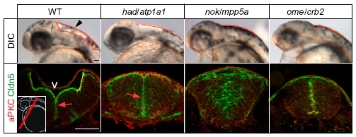
Fig. S4 Phenotypic comparison of brain ventricle expansion defects in different zebrafish ion pump Atp1a1-deficient or cell polarity mutants. Arrowhead indicates location of the hindbrain ventricle in a WT embryo at 30 hpf. Hindbrain ventricular shapes are outlined in red. Sections of immunohistochemical stainings of the brain neuroepithelium show that Cldn5a colocalizes apically together with atypical protein kinase C (aPKC) at the inner ventricular (V) surface at 30 hpf (arrows; Left Inset indicates the cross-sectional plane through the hindbrain region). Localization of Cldn5a and aPKC is affected in nokm520/mpp5a and omem289/crb2 cell polarity mutants. (Scale bar: 50 μm.)
Image
Figure Caption
Acknowledgments
This image is the copyrighted work of the attributed author or publisher, and
ZFIN has permission only to display this image to its users.
Additional permissions should be obtained from the applicable author or publisher of the image.
Open Access.
Full text @ Proc. Natl. Acad. Sci. USA

