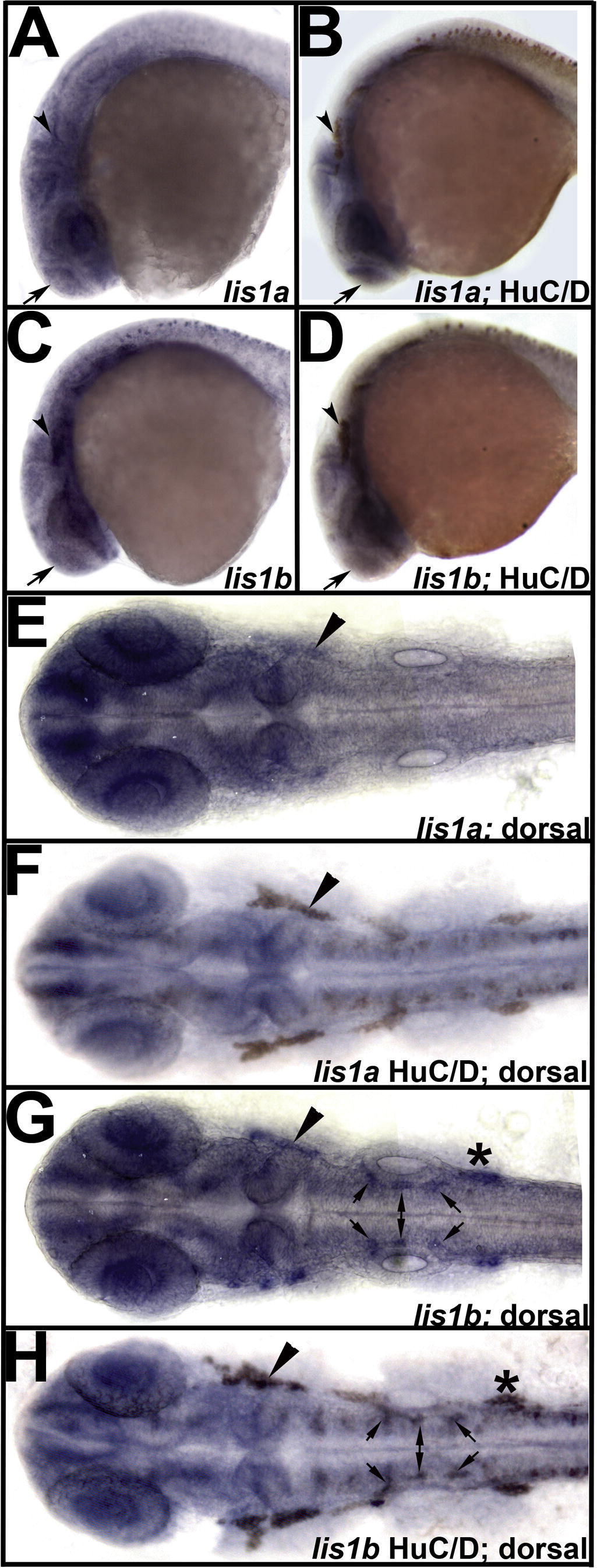
Fig. 8 Immunohistochemical double labeling confirms expression of the LIS1 orthologs in the neurons of the developing central and peripheral neural structures. (A) lis1a expression at 24 hpf is localized to the zebrafish peripheral and central nervous system including the trigeminal ganglion (arrowhead) and forebrain (arrow). (B) This tissue was confirmed as neural by double labeling with an antibody specific for newly matured neurons (HuC/D). (C and D) Similarly, expression of lis1b in the peripheral (arrowhead) and central (arrow) nervous system was confirmed with double labeling for HuC/D. These views are lateral with anterior to the left. (E–H) Dorsal views of 24 hpf embryos further confirm the double labeling of structures for these two orthologs. (E,F) lis1a is expressed highly in the developing forebrain and trigeminal ganglion (arrowhead) at this stage. (G and H) Similarly, lis1b is expressed in the developing forebrain and trigeminal ganglion (arrowhead). Additionally, these dorsal views show strong lis1b expression in other cranial ganglia (*) and punctate groups of neurons in the developing hindbrain (small arrows). These additional areas of expression and the previously described expression in Rohon-Beard sensory neurons are unique for this ortholog.
Reprinted from Gene expression patterns : GEP, 10(1), Drerup, C.M., Wiora, H.M., and Morris, J.A., Characterization of the overlapping expression patterns of the zebrafish LIS1 orthologs, 75-85, Copyright (2010) with permission from Elsevier. Full text @ Gene Expr. Patterns

