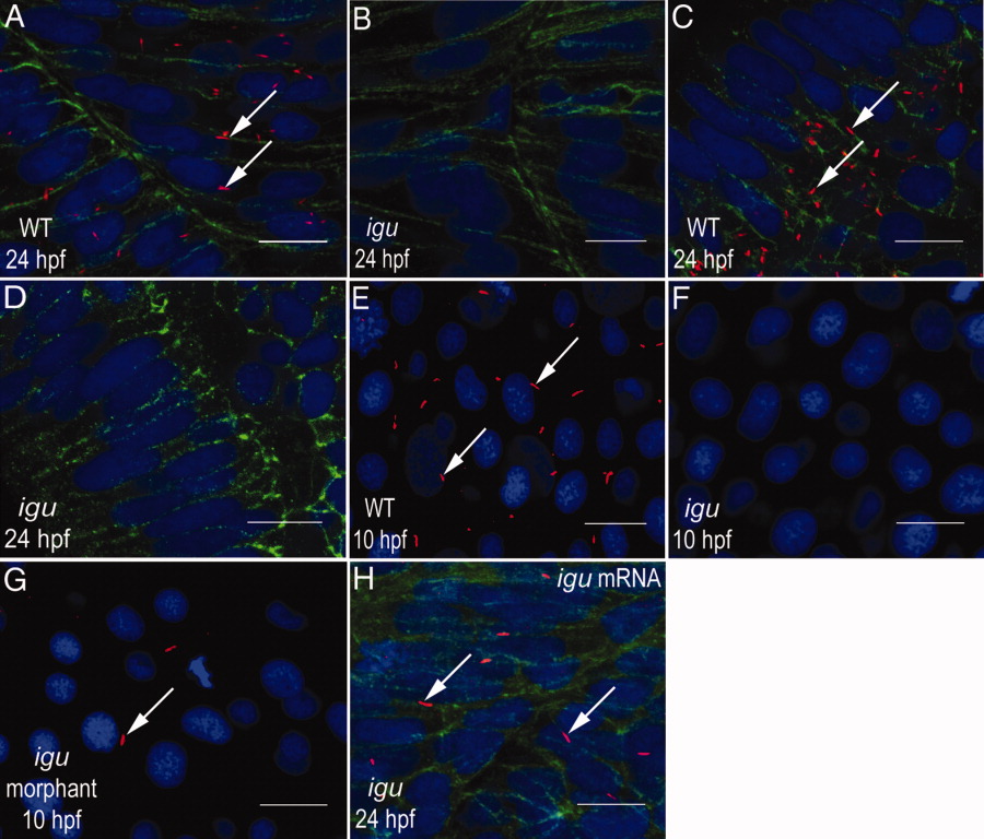
Fig. 1 Igu is essential for primary cilia formation. A: Wild-type myotome showing abundant primary cilia (arrows) on the surface of muscle cells. B: Myotome of an igu mutant embryo showing complete absence of primary cilia. C: Primary cilia (arrows) in differentiating retinal cells of a wild-type embryo. D: Lack of primary cilia from the retina of an igu mutant. E: Primary cilia (arrows) in paraxial mesodermal cells of a wild-type embryo. F: Absence of primary cilia from paraxial mesodermal cells of an igu mutant embryo. G: Severe reduction in numbers of primary cilia (arrow) from paraxial mesodermal cells of an igu morphant embryo. H: An igu mutant embryo injected with igu mRNA showing rescue of the primary cilia (arrows) in muscle cells. Axonemes of primary cilia were visualized with anti-acetylated tubulin antibodies (red), anti-βcatenin antibodies were used to highlight cell membranes (green; A-D and H), and nuclei were visualized with DAPI (blue; 4′,6-diamidine-2-phenylidole-dihydrochloride). In A-D and H, embryos are depicted with anterior to the left and dorsal to the top; E-G show dorsal views of the paraxial mesodermal cells. Scale bars = 10 μm.
Image
Figure Caption
Figure Data
Acknowledgments
This image is the copyrighted work of the attributed author or publisher, and
ZFIN has permission only to display this image to its users.
Additional permissions should be obtained from the applicable author or publisher of the image.
Full text @ Dev. Dyn.

