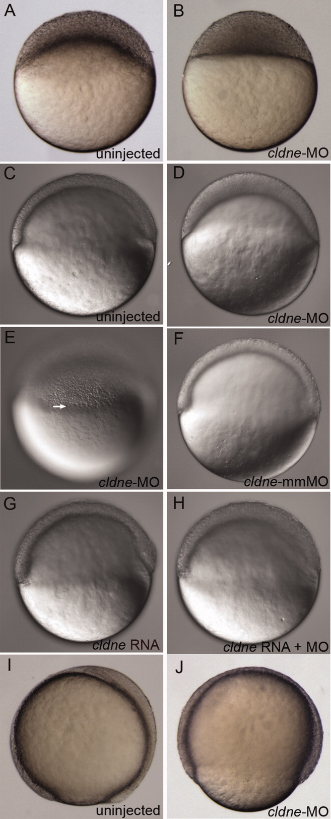
Fig. 2 Embryos injected with cldne-morpholino oligonucleotide (MO) exhibit epiboly delays. Lateral views of live embryos, injected construct indicated in lower right. A,B: Uninjected embryo at 30% epiboly (A), time matched cldne-MO injected embryo at sphere stage (B). C-E: Uninjected embryo at shield stage (C), time matched cldne-MO injected embryo at 40% epiboly (D), same embryo as in D (E), arrow indicates the yolk syncytial layer (YSL) nuclei, which have compacted normally. F: Shield stage embryo injected with mismatch morpholino. G: Shield stage embryo injected with cldne RNA. H: Rescued embryo injected with cldne RNA and cldne-MO. I,J: Uninjected embryo at 90% epiboly stage (I), time matched cldne-MO injected embryo at 70% epiboly (J).
Image
Figure Caption
Figure Data
Acknowledgments
This image is the copyrighted work of the attributed author or publisher, and
ZFIN has permission only to display this image to its users.
Additional permissions should be obtained from the applicable author or publisher of the image.
Full text @ Dev. Dyn.

