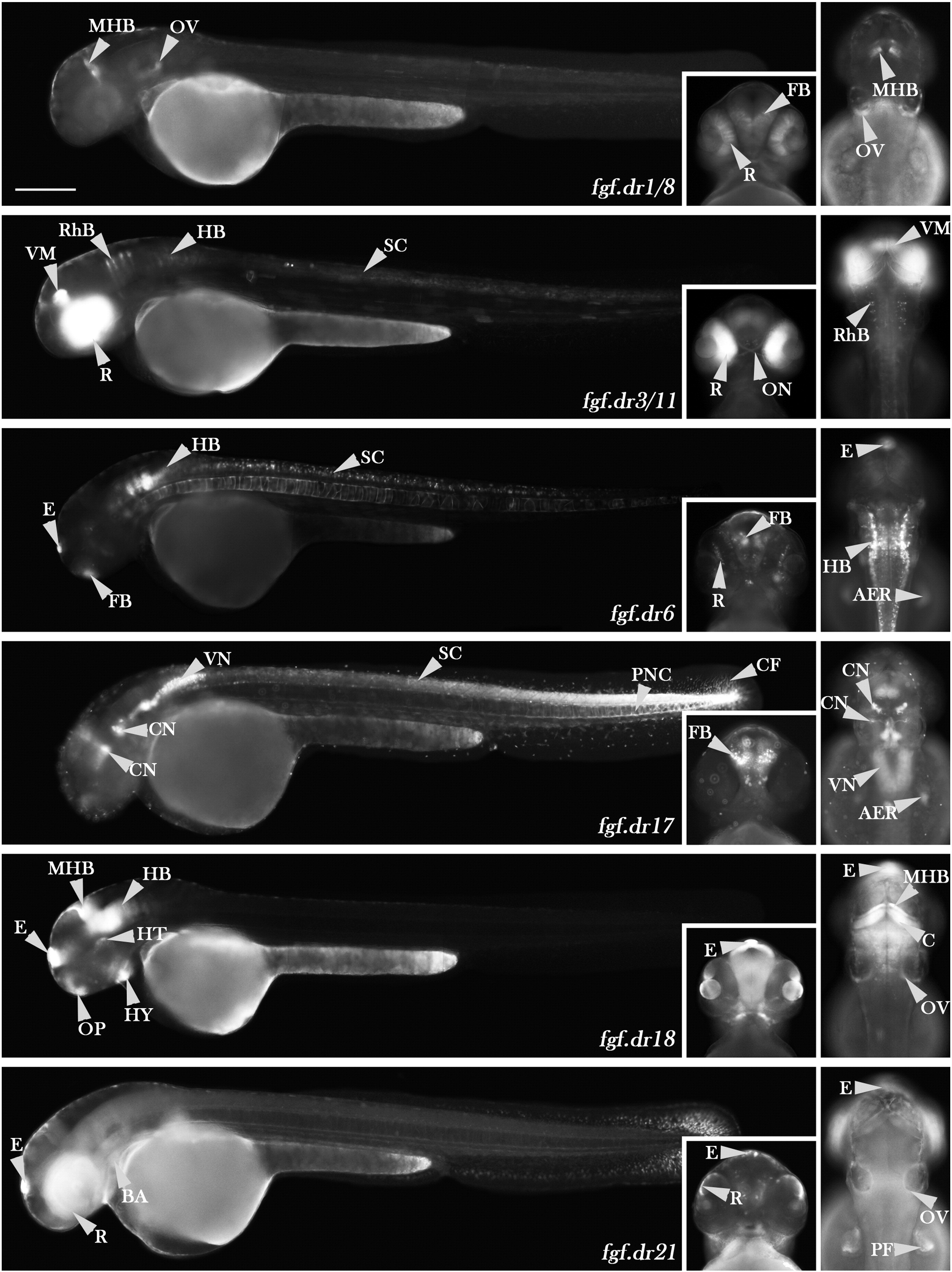
Fig. 4 Reporter gene expression patterns directed by six elements. Element fgf.dr1/8 is active in the midbrain–hindbrain boundary, forebrain, otic vesicle and retina. Element fgf.dr3/11 drove expression in the retina, optic nuclei, ventral midbrain spinal cord and in the rhombomeres. Element fgf.dr6 showed activity in the epiphysis, hindbrain, forebrain, retina, apical ectodermal ridge and spinal cord. Element fgf.dr17 drove GFP expression in cranial motor neurons, vagal nerve, in the hindbrain, apical ectodermal ridge, in the posterior spinal cord and posterior notochord and strongly in the tailbud and caudal fin. Element fgf.dr18 drove expression in all anterior fgf8a expressing structures: in the midbrain–hindbrain boundary, cerebellum, epiphysis, hypothalamus, forebrain, hindbrain, otic vesicle and hyoid cartilage. Element fgf.dr21 drove GFP expression in epiphysis, branchial arches, retina, otic vesicle and pectoral fin. Abbreviations: (AER) apical ectodermal ridge, (BA) branchial arches, (CF) caudal fin, (C) cerebellum, (CN) cranial motor neurons, (E) epiphysis, (FB) forebrain, (HB) hindbrain, (HT) hypothalamus, (HY) hyoid, (MHB) midbrain–hindbrain boundary, (OV) otic vesicle, (ON) optic nerve, (PF) pectoral fin, (PNC) posterior notochord, (R) retina, (RhB) rhombomere boundaries, (SC) spinal cord, (VM) ventral midbrain, (VN) vagal nuclei. Scale bars = 250 μm.
Reprinted from Developmental Biology, 336(2), Komisarczuk, A.Z., Kawakami, K., and Becker, T.S., Cis-regulation and chromosomal rearrangement of the fgf8 locus after the Teleost/tetrapod split, 301-312, Copyright (2009) with permission from Elsevier. Full text @ Dev. Biol.

