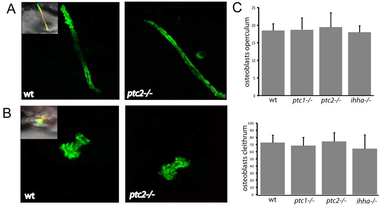
Fig. 2 Mutants with altered Hh signalling show no differences in the number of osterix-positive cells in dermal bone elements. (A) Confocal stacks of the cleithra of osterix-nuclear GFP fish at 4 dpf. Inset image on left is a merged image of Tg(osx:nuGFP) (green), Alazarin Red and brightfield images. (B) Confocal stacks of the operculum of osterix-nuclear GFP fish at 4 dpf. Inset image on left is a merged image of Tg(osx:nuGFP) (green), Alazarin Red and brightfield images. (C) Quantification of the number of osterix-positive nuclei in the cleithrum (below) and operculum (above) at 3 and 4 dpf. Data is shown as mean±s.d. taken of at least four fish per genotype. *P<0.05 versus wild type. wt, wild type.
Image
Figure Caption
Figure Data
Acknowledgments
This image is the copyrighted work of the attributed author or publisher, and
ZFIN has permission only to display this image to its users.
Additional permissions should be obtained from the applicable author or publisher of the image.
Full text @ Development

