
|
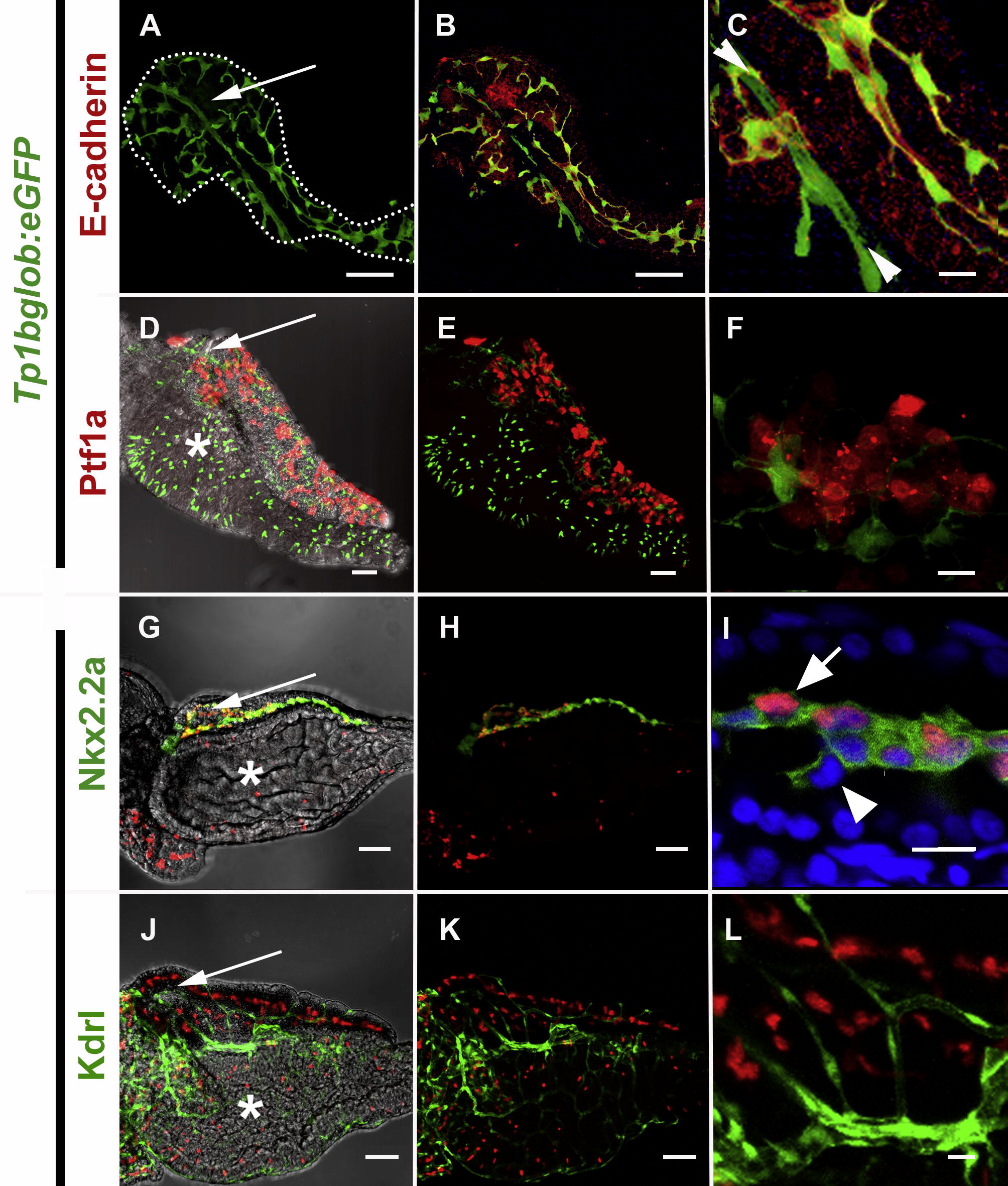
Fig. 3 The localization of pancreatic Notch-responsive cells in the 5 dpf zebrafish pancreas as imaged by confocal microscopy. (A, B, D, E, G, H, J, K) Low power images to see the whole developing organ, outlined (white dots) in (A) (scale bars = 50 μM). (D, G, J) Brightfield merged images allows visualization of pancreas oriented so that the pancreas lies on top of the intestinal bulb (white *) with the principal islet (white →) and head of the pancreas on the left, and the tail of the pancreas on right. (C, F, I, L) High magnification shows detailed cellular structure (scale bars = 10 μM). (A–F) Images of micro-dissected pancreata from Tg(Tp1bglob:eGFP)um14 larvae; PNCs can be visualized as green due to cytoplasmic GFP. GFP positive PNCs can be detected through out the pancreas (A) and immunofluorescent staining to detect E-cadherin (red) indicates vast majority of PNCs are epithelial in nature (B and C). At higher mag. (C), Notch-responsive cells that are E-cad negative can also be seen (►◄). These cells represent endothelium of the early pancreatic, arterial blood supply. (D–F) Tg(ptf1a:Gal4VP16)jh16; Tg(T2KUAS:nfsB-mCherry)jh17 larvae express red fluorescence in a mosaic fashion in the developing exocrine pancreas. The domain of ptf1a driven red fluorescence is exclusive of PNCs. (G–L) Images of micro-dissected pancreata from Tg(T2KTp1bglob:hmgb1-mCherry)jh11, larvae; nuclei in PNCs can be visualized in red. (G–I) Tg(-3.5kbnkx2.2:GFP)ia3 larva expressing green fluorescence in a cell type previously reported to be pancreatic ducts. (I) Some but not all GFP expressing cells are also responding to Notch-activity. (J–L) Tg(kdrlG-RCFP)zn1 larva expressing green fluorescence in the vasculature. With the exception of the same artery imaged in (C), all endothelium is Notch non-responsive.
Reprinted from Mechanisms of Development, 126(10), Parsons, M.J., Pisharath, H., Yusuff, S., Moore, J.C., Siekmann, A.F., Lawson, N., and Leach, S.D., Notch-responsive cells initiate the secondary transition in larval zebrafish pancreas, 898-912, Copyright (2009) with permission from Elsevier. Full text @ Mech. Dev.