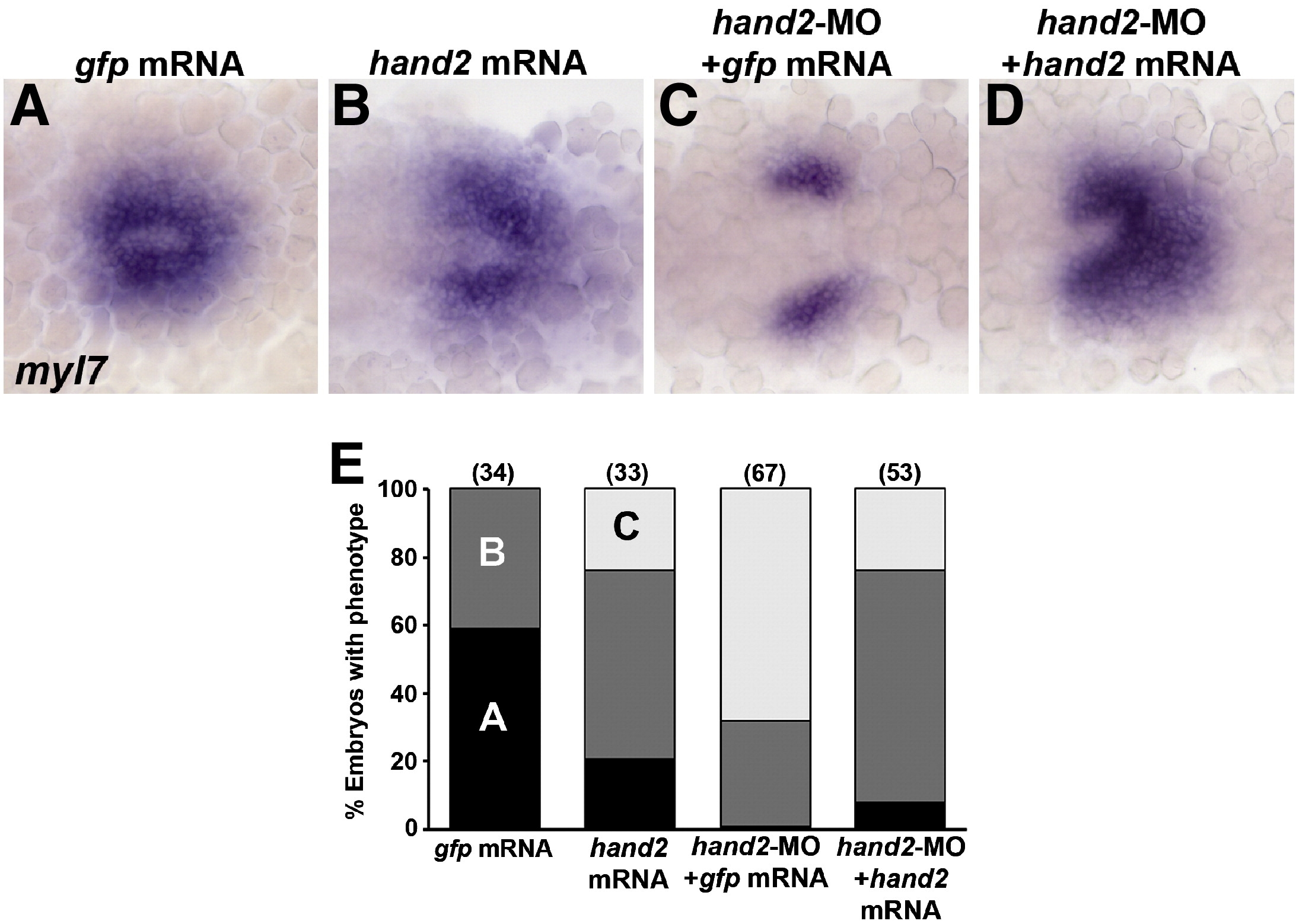
Fig. S1 hand2 RNA rescues myl7 expression in hand2-MO embryos. (A–D) RNA in situ expression of myl7 at the 20s stage in embryos injected with (A) control gfp mRNA, (B) hand2 mRNA, (C), hand2-MO and gfp mRNA, or (D) hand2-MO and hand2 mRNA. Embryos are shown in dorsal view, anterior towards the left. (A) In 20s control embryos, heart precursors form a cone on the embryonic midline. (B) Embryos injected with hand2 mRNA have about wild-type numbers of myl7-expressing cells but heart morphogenesis and cone formation is slightly impaired. (C) Embryos injected with hand2-MO have reduced lateral myl7 domains with defective midline migration. (D) hand2 mRNA largely rescues the hand2-MO phenotype, although most embryos show some impaired morphogenesis. (E) Quantification of the rescue data. Numbers in parentheses denote numbers of embryos. The phenotypic classes correspond to the images shown in panels A–C.
Reprinted from Developmental Biology, 333(2), Maves, L., Tyler, A., Moens, C.B., and Tapscott, S.J., Pbx acts with Hand2 in early myocardial differentiation, 409-418, Copyright (2009) with permission from Elsevier. Full text @ Dev. Biol.

