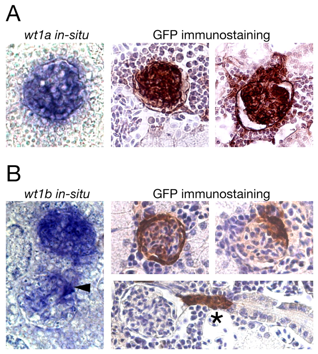
Fig. 3 GFP expression in the mesonephros of adult transgenic zebrafish recapitulates expression of wt1 paralogs. (A,B) In situ hybridization for wt1a (A) and wt1b (B) on sections of wild-type mesonephros (left) and GFP immunostainings on sections of wt1a::GFP line 1 (A) and wt1b::GFP line 1 (B) mesonephros (right) are shown. The kidneys were taken from 4- to 6-month old wild-type and transgenic zebrafish. In the immunostainings, cell nuclei are stained blue (Hematoxylin counterstaining) and GFP-positive cells are brown. Arrowhead marks a glomerulus in which only a subset of cells is labeled, asterisk denotes a GFP-positive neck region.
Image
Figure Caption
Acknowledgments
This image is the copyrighted work of the attributed author or publisher, and
ZFIN has permission only to display this image to its users.
Additional permissions should be obtained from the applicable author or publisher of the image.
Full text @ Development

