
Image
|
Figure Caption
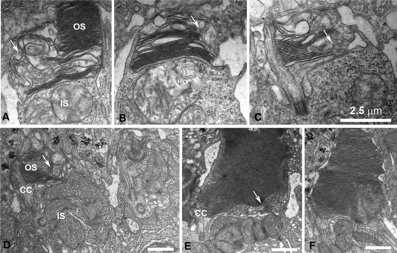
Fig. 8 Disruption of the outer segment (OS) structure in cones expressing DNKIF17. A-D: Electron microscopy (EM) views of cones at the retinal periphery at 5 days after injection of Ta-CP driving DNKIF17. Note disrupted OS structure with failure to complete disc edges as shown by arrows. E: EM view of a cone in the central retina accumulating vesicular membranes within the base of the OS (arrows) opposite the cilium (CC). F: Normal rod photoreceptor. Scale bars = 2.5 μm in C (applies to A-C).
Acknowledgments
This image is the copyrighted work of the attributed author or publisher, and
ZFIN has permission only to display this image to its users.
Additional permissions should be obtained from the applicable author or publisher of the image.
Full text @ Dev. Dyn.