
|
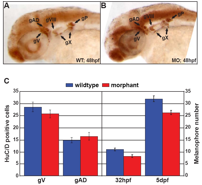
Fig. S7 Disc1 morphants had normal peripheral neuronal development but slightly delayed pigmentation. (A,B) HuC/D immunohistochemistry at 48 hpf in wild type and Disc1 morphants demonstrated identical staining, indicating that Disc1 knockdown does not affect peripheral neuronal development. Lateral views with anterior to the left. (C) There is no difference in HuC/D-positive cell number between wild-type and Disc1 morphant zebrafish in the trigeminal ganglion (gV), which is partially CNC derived. For comparison, the number of HuC/D-positive cells in the anterior lateral line ganglion (gAD), which is derived from placodal tissue, was also counted. In addition, the melanocyte population in the head (anterior to the midbrain-hindbrain boundary) was slightly developmentally delayed in Disc1 morphants but remained comparable to that of wild-type counterparts.